. 2009 Apr 6;185(1):59-66.
doi: 10.1083/jcb.200807073. Epub 2009 Mar 30.

Strabismus regulates asymmetric cell divisions and cell fate determination in the mouse brain

Affiliations

Strabismus regulates asymmetric cell divisions and cell fate determination in the mouse brain

Blue B Lake et al. J Cell Biol. .

Abstract

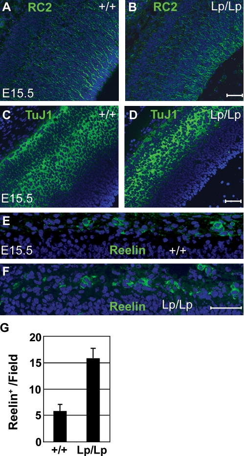
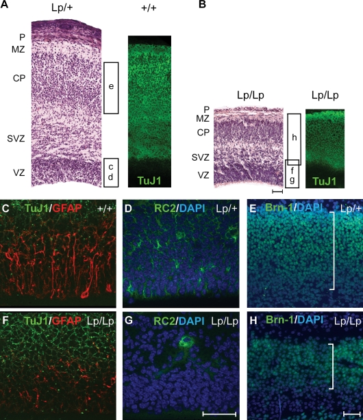
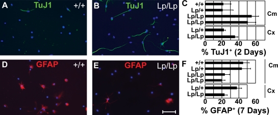
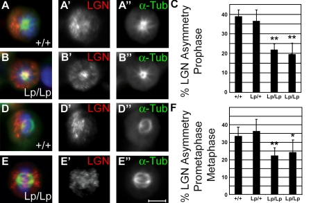
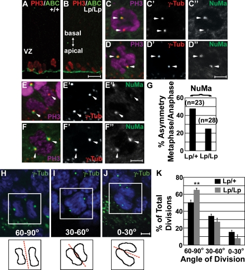
The planar cell polarity (PCP) pathway organizes the cytoskeleton and polarizes cells within embryonic tissue. We investigate the relationship between PCP signaling and cell fate determination during asymmetric division of neural progenitors (NPs) in mouse embryos. The cortex of Lp/Lp (Loop-tail) mice deficient in the essential PCP mediator Vangl2, homologue of Drosophila melanogaster Strabismus (Stbm), revealed precocious differentiation of neural progenitors into early-born neurons at the expense of late-born neurons and glia. Although Lp/Lp NPs were easily maintained in vitro, they showed premature differentiation and loss of asymmetric distribution of Leu-Gly-Asn-enriched protein (LGN)/partner of inscuteable (Pins), a regulator of mitotic spindle orientation. Furthermore, we observed a decreased frequency in asymmetric distribution of the LGN target nuclear mitotic apparatus protein (NuMa) in Lp/Lp cortical progenitors in vivo. This was accompanied by an increase in the number of vertical cleavage planes typically associated with equal daughter cell identities. These findings suggest that Stbm/Vangl2 functions to maintain cortical progenitors and regulates mitotic spindle orientation during asymmetric divisions in the vertebrate brain.


Figures

Figure 1.
Figure 1.
Increased number of Reelin-positive neurons in E15.5 Lp/Lp cerebral cortices. (A–F) Coronal sections of Lp/Lp or wild-type cortices (E15.5) were stained for RC2 (A and B), TuJ1 (C and D), Reelin (E and F, green), and DAPI (blue). (G) The number of Reelin-positive neurons is increased in three different Lp/Lp embryos (15.8 ± 1.9 per field, scored over four 110-µm-wide fields; F) as compared with wild-type mice (5.8 ± 1.3 per field; E). The total number of DAPI-positive nuclei in the marginal layer (scored over six 100-µm-wide fields per genotype) is 72.2 ± 7.4 for Lp/Lp and 61.2 ± 8.3 for wild-type littermates. Error bars indicate mean ± SD. Bars, 50 µm.
Figure 2.
Figure 2.
Precocious differentiation of NPs in Lp/Lp mice. (A–H) Coronal sections of Lp/Lp cortices at E18.5 are shown. They are reduced in size and show premature depletion of progenitors. Histological analysis of E18.5 neocortex, hematoxylin/eosin staining, and the corresponding TuJ1 staining (right) are shown for wild-type and Lp/+ (A) and Lp/Lp (B) cortices. The respective positions of the pial layer (P), marginal zone (MZ; layer I), cortical plate (CP; layers II–VI), sub-VZ (SVZ), and VZ are shown. The corresponding regions examined in C–H are also shown. The astrocytes marked by GFAP (C and F), radial glia progenitors marked by RC2 (D and G), and late-born outer layer neurons marked by Brn-1 (E and H) were all significantly reduced in Lp/Lp mice. Brackets in E and H represent the relative size of the Brn-1 layer. Bars, 50 µm.
Figure 3.
Figure 3.
Precocious differentiation of Lp/Lp NPs in vitro. Progenitors derived from the E18.5 cerebellum (Cm) or E14.5 cortex (Cx) were obtained from Lp/Lp mice and compared with NPs obtained from Lp/+ and/or +/+ littermates. Cerebellar progenitors were also obtained and compared between two different Lp/Lp mice. Dissociated cells were cultured in vitro under conditions that promote sequential neuronal then astrocytic differentiation. (A–C) In the absence of bFGF/EGF for 2 d, NPs formed TuJ1-positive neurons. (D–F) Addition of 2% serum to these cells at day 3 and culture for four additional days allowed astrocyte differentiation that was scored by GFAP staining. (A, B, D, and E) Images shown are for cerebellar progenitors. Frequencies of TuJ1+ (C) and GFAP+ (F) cells were scored per total number of DAPI-positive cells. Error bars indicate mean ± SD. Bar, 50 µm.
Figure 4.
Figure 4.
Reduced number of asymmetric divisions in Lp/Lp progenitors. NPs were allowed to differentiate for 24 h in the bFGF/B27 medium. (A, B, D, and E) 0.4 µg/ml nocodazole was added for the last 6.5 h and removed 0.5 h before cells were fixed and costained with antibodies against α-tubulin, LGN, and DAPI. Prophase (A and B) and prometaphase/metaphase (D and E) cells were identified by α-tubulin localization and exhibited either asymmetric (A–A″ and D–D″) or symmetric (B–B″ and E–E″) localization of LGN. The total number of cell divisions with LGN asymmetry (C and F) was scored at prophase (Lp/Lp embryo 1, n = 257; Lp/Lp embryo 2, n = 294; Lp/+, n= 228; +/+, n = 207) and prometaphase/metaphase (Lp/Lp embryo 1, n = 416; Lp/Lp embryo 2, n = 445; Lp/+, n = 351; +/+, n = 331). Statistical significance was determined against the Lp/+ condition by a standard two-tailed Student's t test (*, P < 0.05; **, P < 0.01). Error bars indicate mean ± SD. Bar, 5 µm.
Figure 5.
Figure 5.
Asymmetric divisions of NPs in the developing cortex. (A and B) Coronal cryosections of E14.5 Lp/Lp (n = 3) and Lp/+ (n = 3) forebrains were stained for phosphohistone H3 (PH3) to identify dividing VZ progenitors. Apical surfaces facing the lateral ventricle are indicated by nonphosphorylated (activated) β-catenin (ABC) staining. (C–F) Only cells adjacent to the lateral ventricle were analyzed and shown with their apical surfaces oriented down. Asymmetric (C–C″ and E–E″) and symmetric (D–D″ and F–F″) centrosomal localization of NuMa at prometaphase (C–D) and anaphase (E–F). Metaphase to anaphase NuMa asymmetries are summarized in G. Images shown are of Lp/+ (C) and Lp/Lp (D–F) cortices. Arrows indicate centrosomes. (H–J) Cleavage plane orientation with respect to the apical surface was scored as described in Materials and methods and is presented in three broad categories: 60–90°, 30–60°, and 0–30°. Representative images are shown. Boxed regions indicate telophase nuclei represented graphically. (K) Comparison of cleavage plane orientation during anaphase/telophase of cortical cell divisions in Lp/+ (n = 172) and Lp/Lp (n = 112) embryos (three for each genotype). Statistical significance was determined by a standard two-tailed Student's t test (**, P < 0.01). Error bars indicate mean ± SD. Bars: (B) 25 µm; (D″, F″, and J) 5 µm.

References

    1. Bellaiche Y., Beaudoin-Massiani O., Stuttem I., Schweisguth F. 2004. The planar cell polarity protein Strabismus promotes Pins anterior localization during asymmetric division of sensory organ precursor cells in Drosophila.Development. 131:469–478 - PubMed
    1. Betschinger J., Knoblich J.A. 2004. Dare to be different: asymmetric cell division in Drosophila, C. elegans and vertebrates.Curr. Biol. 14:R674–R685 - PubMed
    1. Blumer J.B., Chandler L.J., Lanier S.M. 2002. Expression analysis and subcellular distribution of the two G-protein regulators AGS3 and LGN indicate distinct functionality. Localization of LGN to the midbody during cytokinesis.J. Biol. Chem. 277:15897–15903 - PubMed
    1. Bond J., Roberts E., Mochida G.H., Hampshire D.J., Scott S., Askham J.M., Springell K., Mahadevan M., Crow Y.J., Markham A.F., et al. 2002. ASPM is a major determinant of cerebral cortical size.Nat. Genet. 32:316–320 - PubMed
    1. Brott B.K., Sokol S.Y. 2005. A vertebrate homolog of the cell cycle regulator Dbf4 is an inhibitor of Wnt signaling required for heart development.Dev. Cell. 8:703–715 - PubMed

Publication types