. 2009;4(3):e5070.
doi: 10.1371/journal.pone.0005070. Epub 2009 Mar 31.

A variant mimicking hyperphosphorylated 4E-BP inhibits protein synthesis in a sea urchin cell-free, cap-dependent translation system

Affiliations

A variant mimicking hyperphosphorylated 4E-BP inhibits protein synthesis in a sea urchin cell-free, cap-dependent translation system

Nathalie Oulhen et al. PLoS One. 2009.

Abstract

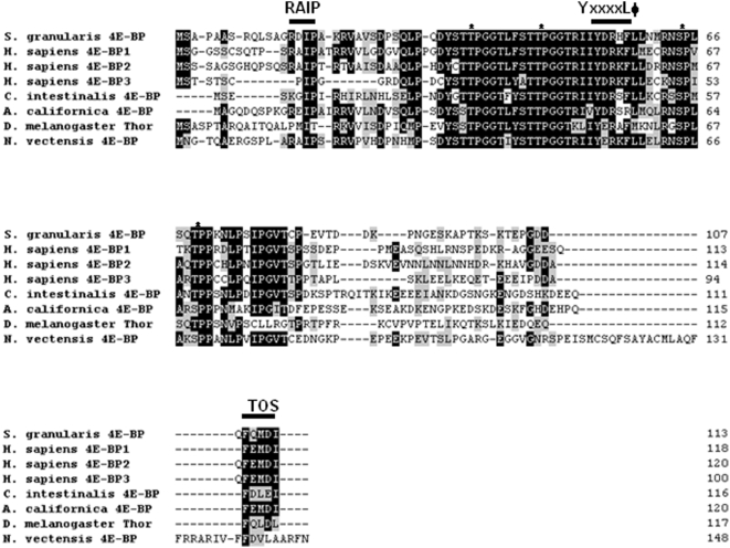
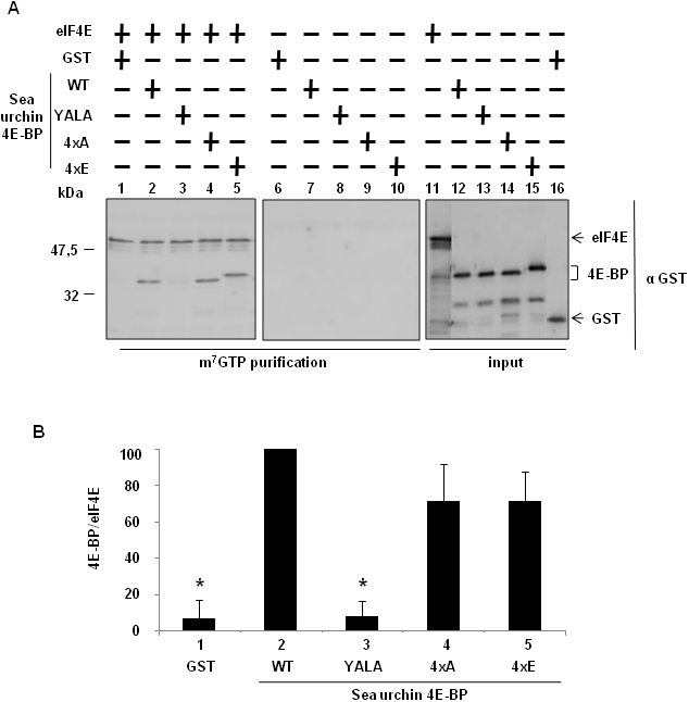
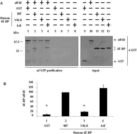
Background: 4E-BP is a translational inhibitor that binds to eIF4E to repress cap-dependent translation initiation. This critical protein:protein interaction is regulated by the phosphorylation of 4E-BP. Hypophosphorylated 4E-BP binds to eIF4E and inhibits cap-dependent translation, whereas hyperphosphorylated forms do not. While three 4E-BP proteins exist in mammals, only one gene encoding for 4E-BP is present in the sea urchin genome. The protein product has a highly conserved core domain containing the eIF4E-binding domain motif (YxxxxLPhi) and four of the regulatory phosphorylation sites.

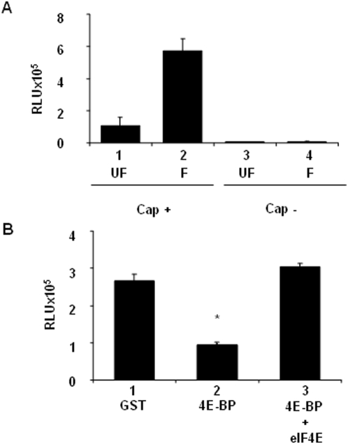
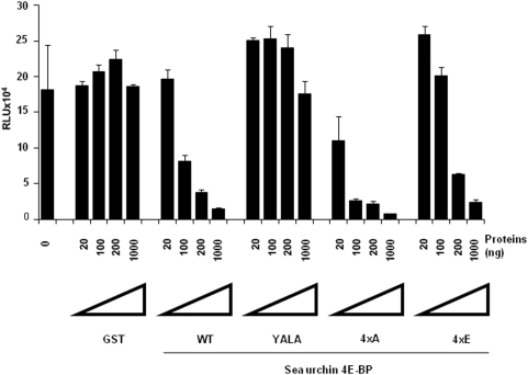
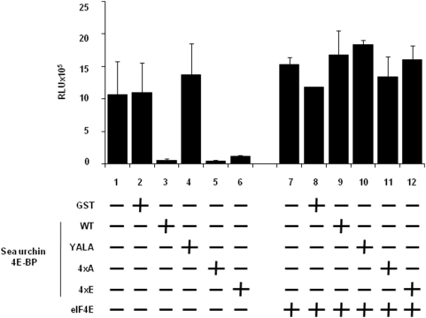
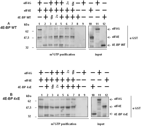
Methodology/principal findings: Using a sea urchin cell-free cap-dependent translation system prepared from fertilized eggs, we provide the first direct evidence that the sea urchin 4E-BP inhibits cap-dependent translation. We show here that a sea urchin 4E-BP variant, mimicking phosphorylation on four core residues required to abrogate binding to eIF4E, surprisingly maintains physical association to eIF4E and inhibits protein synthesis.

Conclusions/significance: Here, we examine the involvement of the evolutionarily conserved core domain and phosphorylation sites of sea urchin 4E-BP in the regulation of eIF4E-binding. These studies primarily demonstrate the conserved activity of the 4E-BP translational repressor and the importance of the eIF4E-binding domain in sea urchin. We also show that a variant mimicking hyperphosphorylation of the four regulatory phosphorylation sites common to sea urchin and human 4E-BP is not sufficient for release from eIF4E and translation promotion. Therefore, our results suggest that there are additional mechanisms to that of phosphorylation at the four critical sites of 4E-BP that are required to disrupt binding to eIF4E.


Conflict of interest statement

Competing Interests: The authors have declared that no competing interests exist.

Figures

Figure 1
Figure 1. Amino acid sequences of 4E-BP proteins.
Sg4E-BP protein was predicted from the cDNA and aligned with the three human 4E-BPs, Ciona intestinalis 4E-BP, Drosophila melanogaster Thor, Aplysia californica 4E-BP and Nematostella vectensis 4E-BP. Accession numbers of human proteins are: 4E-BP1 (NP_004086), 4E-BP2 (Q13542), 4E-BP3 (NP_003723) on NCBI. The Accession number is NP_477295 for Drosophila melanogaster (NCBI), 299279 for Ciona intestinalis (http://genome.jgi-psf.org/Cioin2/) and SB_47700 for Nematostella vectensis (http://www.stellabase.org/). The Aplysia californica sequence 4E-BP was obtained from . The four residues known to be phosphorylated on human 4E-BPs and conserved on sea urchin are indicated by stars. Identical and conserved amino acid residues are on black and grey background, respectively. The common eIF4E motif indicated by YxxxxLΦ (where x is any amino acid and Φ is an hydrophobic residue), the RAIP motif and the TOS (TOR signalling) site are denoted with a line above.
Figure 2
Figure 2. Recombinant GST-Sg4E-BP (4E-BP) interacts with endogenous SgIF4E isoforms in sea urchin extracts.
After incubation of the GST alone (lanes 1–2) or the GST-Sg4E-BP protein (lanes 3–4) in extract prepared from unfertilized eggs (UF, lanes 1 and 3) or from 60 minutes post-fertilization embryos (F, lanes 2 and 4), proteins were affinity purified using Gluthatione Sepharose 4B beads, resolved by 15% SDS-PAGE, analysed by immunoblotting and detected by chemiluminescence using an anti-GST antibody (top and intermediate panels) or anti-eIF4E antibody (bottom panels) as described in Materials and Methods. SgIF4E that co-purified with GST-Sg4E-BP (lanes 3 and 4) was compared with the endogenous SgIF4E detected in 10 µg of total protein extracts (corresponding to 0,5% of the volume used for the purification) loaded separately (lanes 5–6).
Figure 3
Figure 3. Sg4E-BP inhibits protein synthesis activity in sea urchin cell-free cap-dependent translation system.
Cell-free translation system from unfertilized eggs (UF) and 30 minutes post-fertilization embryos (F) were prepared as described in Materials and Methods. (A) Renilla Luciferase activity was measured after addition of capped (Cap+) or uncapped (Cap−) mRNA. Error bars represent the standard deviation (s.d.) of duplicates. (B) Sg4E-BP inhibits cap dependent translation activity and this inhibition is rescued by eIF4E. 100 ng of GST (lane 1), 100 ng of GST-Sg4E-BP alone (4E-BP, lane 2) or preincubated 5 min with 250 ng of GST-mIF4E (4E-BP+eIF4E, lane 3) were added to the fertilized cell free translation system and Luciferase activity was measured as described in Materials and Methods after addition of a capped mRNA encoding Renilla Luciferase. Error bars represent the standard deviation (s.d.) of duplicates. Significance was assessed using Fisher's F-test and Student's t-test. *P<0.005, significant difference between 4E-BP and GST, and between 4E-BP and 4E-BP+eIF4E.
Figure 4
Figure 4. The Sg4E-BP variant mimicking hyperphosphorylation inhibits protein synthesis in sea urchin cell-free cap-dependent translation system.
Different amounts (20; 100; 200 or 1,000 ng) of recombinant proteins (GST, GST-Sg4E-BP WT (WT), GST-Sg4E-BP YALA (YALA), GST-Sg4E-BP 4xA (4xA), GST-Sg4E-BP 4xE (4xE)) were added to the fertilized cell-free translation system and Luciferase activity was measured as described in Materials and Methods after addition of the Cap+mRNA encoding Renilla Luciferase. The Luciferase activity is represented in RLU (Relative Light Units). Error bars represent the standard deviation (s.d.) of duplicates.
Figure 5
Figure 5. Translation inhibition is rescued by GST-mIF4E.
200 ng of GST recombinant proteins (GST, GST-Sg4E-BP WT (WT), GST-Sg4E-BP YALA (YALA), GST-Sg4E-BP 4xA (4xA), GST-Sg4E-BP 4xE (4xE)) were added to the fertilized cell-free translation system with (lanes 8–12) or without (lanes 2–6) a 5 min pre-incubation with 500 ng of GST-mIF4E (eIF4E) and Luciferase activity was measured as described in Materials and Methods after addition of a cap mRNA encoding Renilla Luciferase. The Luciferase activity is represented in RLU (Relative Light Units). Error bars represent the standard deviation (s.d.) of duplicates.
Figure 6
Figure 6. The variant mimicking hyperphosphorylation of Sg4E-BP also binds to eIF4E.
(A) The S/T-E Sg4E-BP mutated at all four phosphorylation sites (4xE) binds to eIF4E. GST-mIF4E (eIF4E) was incubated with m7GTP sepharose beads (lanes 1–5) and the GST recombinant proteins were added: GST, GST-Sg4E-BP WT (WT), GST-Sg4E-BP YALA (YALA), GST-Sg4E-BP 4xA (4xA), GST-Sg4E-BP 4xE (4xE). Complexes were affinity purified and analysed by Western blot using a GST antibody. Lanes 6–10, GST-mIF4E was omitted as control for binding specificity. Inputs are shown on the right panel (lanes 11–16). They represent 10% of the volume used in the experiment. (B) Binding between the GST recombinant proteins and GST-mIF4E was analysed by quantification of the signals obtained on the Fig 6.A, using Image Quant software. Error bars represent the standard deviation (s.d.) of two experiments. Significance was assessed using Fisher's F-test and Student's t-test. * P<0.01 significant difference between GST or GST-Sg4E-BP YALA with GST-Sg4E-BP WT.
Figure 7
Figure 7. Wild type Sg4E-BP and the S/T-E variant inhibit eIF4E/eIF4G association with the same efficiency.
Wild type (A) and the 4xE variant of Sg4E-BP (B) inhibit eIF4E/eIF4G association. The m7GTP sepharose beads was incubated (lanes 1–7) or not (lanes 8–9) with GST-mIF4E (eIF4E). Then, recombinant proteins GST-Sg4E-BP WT (WT, A) or GST-Sg4E-BP WT 4xE (4xE, B) and GST-SgIF4G (eIF4G) were added separately for lanes 1–2 and lanes 8–9, and together for other lanes (3-4-5-6-7). We used the same amount of GST-Sg4E-BP and GST-SgIF4G in lane 5, a 2-fold ratio in lanes 4 and 6 and a 10-fold ratio in lanes 3 and 7. Proteins were affinity-purified using m7GTP sepharose beads and were analysed by immunoblotting as described in Materials and Methods using an anti-GST antibody. Affinity-purified proteins were compared with the GST-fusion proteins loaded separately (lanes 10–12). Inputs represent 10% of the volume used in the experiment.
Figure 8
Figure 8. The variant mimicking hyperphosphorylation of human 4E-BP does not decrease its association with eIF4E.
(A) The S/T-E human 4E-BP mutated at all four phosphorylation sites (4xE) binds to eIF4E. GST-mIF4E (eIF4E) was incubated with m7GTP sepharose beads (lanes 1–4) and the GST recombinant proteins were added: GST, GST-Hu4E-BP WT (WT), GST-Hu4E-BP YALA (YALA), GST-Hu4E-BP 4xE (4xE). Complexes were affinity purified and analysed by Western blot using a GST antibody. Lanes 5–8, GST-mIF4E was omitted as control for binding specificity. Inputs are shown on the right panel (lanes 9–13), they represent 10% of the volume used in the experiment. (B) Binding between the GST recombinant proteins and GST-mIF4E was analysed by quantification of the signals obtained from duplicates of two independent experiments on the Fig 8.A using Image Quant software. Error bars represent the standard deviation (s.d.) of two experiments. Significance was assessed using Fisher's F-test and Student's t-test. * P<0.01 significant difference between GST or GST-Hu4E-BP YALA with GST-Hu4E-BP WT.

Similar articles

Cited by

References

    1. Mathews MB, Sonenberg N, Hershey JWB. Origins and Principles of Translational Control. In: Mathews MB, Sonenberg N, Hershey JWB, editors. Translational Control in Biology and Medicine. Cold Spring Harbor, NY: Cold Spring Harbor Laboratory Press; 2007.
    1. Davidson EH, Hough-Evans BR, Britten RJ. Molecular biology of the sea urchin embryo. Science. 1982;217:17–26. - PubMed
    1. Epel D. Protein synthesis in sea urchin eggs: a “late” response to fertilization. Proc Natl Acad Sci U S A. 1967;57:899–906. - PMC - PubMed
    1. Epel D. The initiation of development at fertilization. Cell Differ Dev. 1990;29:1–12. - PubMed
    1. Humphreys T. Measurements of messenger RNA entering polysomes upon fertilization of sea urchin eggs. Dev Biol. 1971;26:201–208. - PubMed

Publication types

Substances