Review
. 2009 Jul;66(14):2299-318.
doi: 10.1007/s00018-009-0020-8. Epub 2009 Mar 31.

Amyloid precursor protein and its homologues: a family of proteolysis-dependent receptors

Affiliations
Review

Amyloid precursor protein and its homologues: a family of proteolysis-dependent receptors

Kristin T Jacobsen et al. Cell Mol Life Sci. 2009 Jul.

Abstract

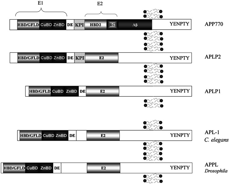
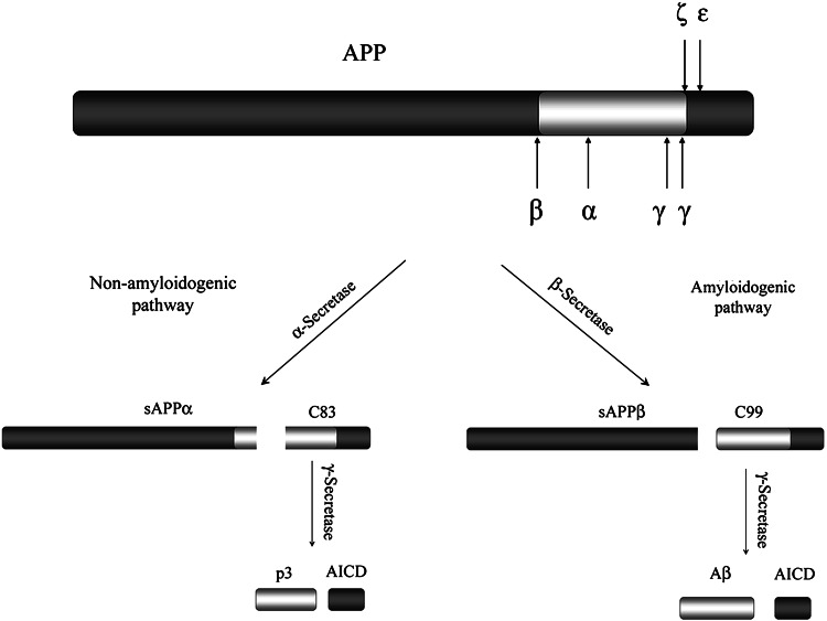
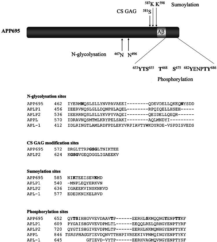
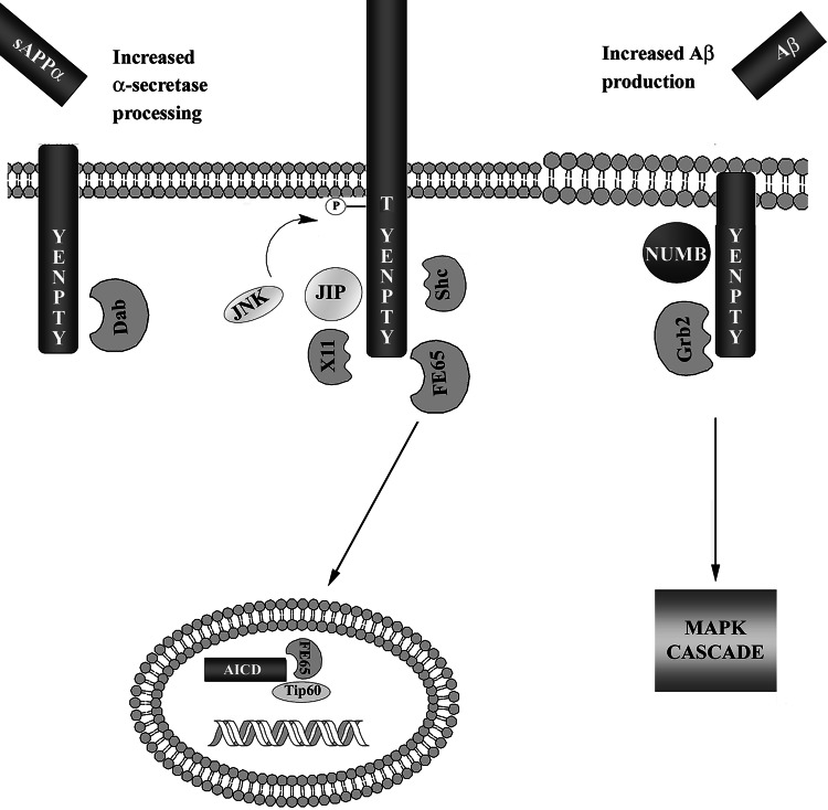
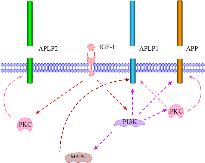
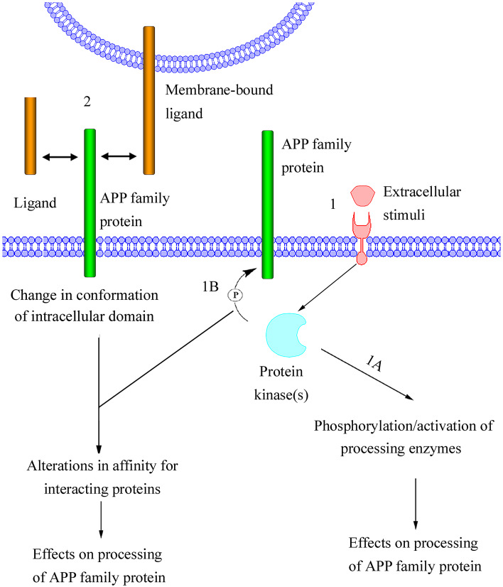
The Alzheimer's amyloid precursor protein (APP) belongs to a conserved gene family that also includes the mammalian APLP1 and APLP2, the Drosophila APPL, and the C. elegans APL-1. The biological function of APP is still not fully clear. However, it is known that the APP family proteins have redundant and partly overlapping functions, which demonstrates the importance of studying all APP family members to gain a more complete picture. When APP was first cloned, it was speculated that it could function as a receptor. This theory has been further substantiated by studies showing that APP and its homologues bind both extracellular ligands and intracellular adaptor proteins. The APP family proteins undergo regulated intramembrane proteolysis (RIP), generating secreted and cytoplasmic fragments that have been ascribed different functions. In this review, we will discuss the APP family with focus on biological functions, binding partners, and regulated processing.


Figures

Fig. 1
Fig. 1
Schematic illustration of the domain organization of APP and its homologues. All members of the APP family contain heparin-binding/growth-factor-like domains (HBD/GFLD), copper- and zinc-binding domains (CuBD and ZnBD), an acidic domain (DE), and a protein interaction motif (YENPTY) in the C-terminal
Fig. 2
Fig. 2
Schematic illustration of APP processing. The major cleavage of APP in the ectoplasmic domain occurs at the α- and β-site, resulting in the secretion of sAPPα or sAPPβ. The remaining C-terminal membrane-bound fragments, C83 or C99, are subsequently cleaved within the transmembrane region by γ-secretase, releasing AICD and p3 or
Fig. 3
Fig. 3
Posttranslational modification sites in APP and its homologues. Schematic illustration of posttranslational modification sites in APP. Note, APP also undergoes O-glycosylation, but the site has still not been determined. Alignment of the APP modification sequences with its homologues. The APP N 467 N-glycosylation site is conserved in all homologues, whereas the APP K 695 sumoylation site is only conserved within the mammalian homologues. Three known in vivo phosphorylation sites (T 668, Y 682, T 686, and Y 687) are conserved in all homologues and one additional site (Y 653) in APLP1 and APLP2. The proteins were aligned using the BLASTp Blossum62 matrix
Fig. 4
Fig. 4
Schematic illustration of proposed interactions between APP and adaptor proteins. APP interacts with several adaptor proteins through the YENPTY motif in its intracellular domain. The interactions between APP and the adaptor proteins affect the metabolism of APP. Dab1 interaction increases α-secretase processing, while Grb2 and Numb increase β-secretase processing. JIP, X11, and Shc stabilize full-length APP. Note that β-secretase cleavage of APP is considered to take place in endosomes or lipid rafts, as illustrated by the different appearance of the membrane. JIP and X11 both regulate the phosphorylation at Thr668 through JNK, thereby regulating the interaction with other adaptor proteins. Grb2 interaction with APP results in activation of the MAPK cascade, similar to Grb2 interaction with tyrosine kinase receptors. The γ-secretase generated fragment AICD has been demonstrated to translocate into the nucleus where it forms a complex with FE65 and Tip60. The complex has been suggested to regulate gene transcription (see the text for references)
Fig. 5
Fig. 5
Schematic illustration of IGF-1-induced signaling resulting in increased processing of the mammalian APP family proteins. IGF-1-induced processing of APP and APLP1 is almost completely dependent on activation of PI3K, but is also affected by PKC activation. This indicates that PKC acts downstream of PI3K. In contrast, IGF-1-induced processing of APLP2 is independent of PI3K activation but dependent on PKC activation, indicating that PKC also is activated independently of PI3K during IGF-1 stimulation. Note that different PKC isoforms could be involved. Only the IGF-1-induced processing of APLP1 was shown to be dependent on MAPK activation. A model is proposed where IGF-1 binds and activates membrane-bound IGF-1 receptors, resulting in activation of PI3K and PKC, independently of each other
Fig. 6
Fig. 6
Proposed model of regulated processing of APP family proteins. In this model, processing is induced either (1) by an extracellular stimuli (e.g., growth factors) or (2) through direct interaction with a soluble ligand (e.g., sAPPα), an extracellular matrix protein (e.g., F-spondin; not shown) or a membrane-bound ligand (e.g., TAG-1, APP, APLP1, or APLP2). The extracellular stimuli activate membrane-bound receptors, resulting in activation of cytoplasmic protein kinases. One possible effect of the activated protein kinases is phosphorylation of proteins involved in cleavage of APP family proteins (1A). This could result in altered activity or intracellular localization of the enzymes, thereby affecting the processing of APP family proteins. Alternatively, the activated protein kinases could phosphorylate APP family proteins (1B). Phosphorylation of the cytosolic domain of APP family proteins would most likely affect the affinity for interacting proteins. These interacting proteins could be adaptor proteins, which regulate APP family protein localization, resulting in altered proximity of APP family proteins to processing enzymes. The phosphorylation of the intracellular domain could also turn APP family proteins into better substrates for enzymatic cleavage. It is likely that the interaction of APP family proteins with soluble or membrane-bound ligands would affect the conformation of the intracellular domain of APP family proteins. In this model, the conformational change, like the extracellular stimuli-induced phosphorylation of APP family proteins is thought to alter the affinity for interacting proteins, thereby affecting the processing of APP family proteins

References

    1. Kang J, Lemaire HG, Unterbeck A, Salbaum JM, Masters CL, Grzeschik KH, Multhaup G, Beyreuther K, Müller-Hill B. The precursor of Alzheimer’s disease amyloid A4 protein resembles a cell-surface receptor. Nature. 1987;325:733–736. doi: 10.1038/325733a0. - DOI - PubMed
    1. Wasco W, Bupp K, Magendantz M, Gusella JF, Tanzi RE, Solomon F. Identification of a mouse brain cDNA that encodes a protein related to the Alzheimer disease-associated amyloid beta protein precursor. Proc Natl Acad Sci USA. 1992;89:10758–10762. doi: 10.1073/pnas.89.22.10758. - DOI - PMC - PubMed
    1. Wasco W, Gurubhagavatula S, Paradis MD, Romano DM, Sisodia SS, Hyman BT, Neve RL, Tanzi RE. Isolation and characterization of APLP2 encoding a homologue of the Alzheimer’s associated amyloid beta protein precursor. Nat Genet. 1993;5:95–100. doi: 10.1038/ng0993-95. - DOI - PubMed
    1. Rosen DR, Martin-Morris L, Luo LQ, White K. A Drosophila gene encoding a protein resembling the human β-amyloid protein precursor. Proc Natl Acad Sci USA. 1989;86:2478–2482. doi: 10.1073/pnas.86.7.2478. - DOI - PMC - PubMed
    1. Daigle I, Li C. apl-1, a Caenorhabditis elegans gene encoding a protein related to the human β-amyloid protein precursor. Proc Natl Acad Sci USA. 1993;90:12045–12049. doi: 10.1073/pnas.90.24.12045. - DOI - PMC - PubMed

MeSH terms

LinkOut - more resources