Review
. 2009 Aug;66(15):2405-26.
doi: 10.1007/s00018-009-0017-3. Epub 2009 Mar 31.

Estrogen and progesterone receptors: from molecular structures to clinical targets

Affiliations
Review

Estrogen and progesterone receptors: from molecular structures to clinical targets

Stephan Ellmann et al. Cell Mol Life Sci. 2009 Aug.

Abstract

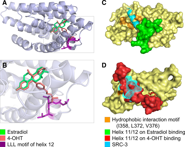
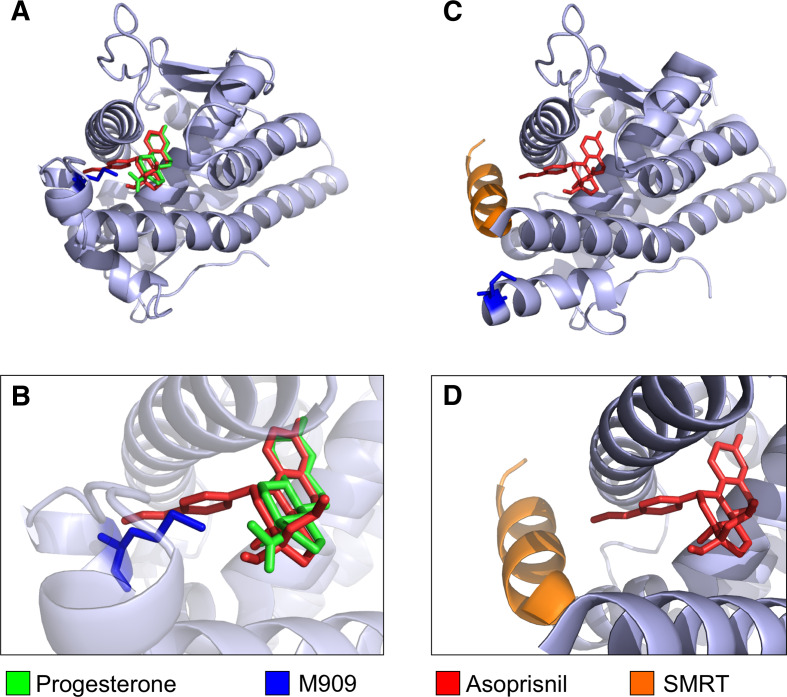
Research involving estrogen and progesterone receptors (ER and PR) have greatly contributed to our understanding of cell signaling and transcriptional regulation. In addition to the classical ER and PR nuclear actions, new signaling pathways have recently been identified due to ER and PR association with cell membranes and signal transduction proteins. Bio-informatics has unveiled how ER and PR recognize their ligands, selective modulators and co-factors, which has helped to implement them as key targets in the treatment of benign and malignant tumors. Knowledge regarding ER and PR is vast and complex; therefore, this review will focus on their isoforms, signaling pathways, co-activators and co-repressors, which lead to target gene regulation. Moreover it will highlight ER and PR involvement in benign and malignant diseases as well as pharmacological substances influencing cell signaling and provide established and new structural insights into the mechanism of activation and inhibition of these receptors.


Figures

Fig. 1
Fig. 1
Phylogenetic analysis of the nuclear receptor superfamily based on human protein sequences. a Schematic shows the 18 nuclear receptor (NR) members and common ancestor (CA). b Unrooted tree showing evolutionary distances between the members of the nuclear receptor superfamily. Distances in both trees correlate directly to evolutionary distances and inversely to sequence identity of the proteins analyzed, including estrogen receptor α and β (ERα/β), glucocorticoid (GR), mineralocorticoid (MR), progesterone (PR), androgen (AR), retinoic acid (RARα/β/γ), retinoid X (RXRα/β/γ), vitamin D (VDR), peroxisome proliferator activated (PPARα/γ/δ) and thyroid receptor (TRα/β). In addition to the 18 NR members several orphan receptors have been described, which are not represented in the schematics. One of these orphan receptor subgroups is called NR3B, where ERRα is most abundantly expressed, followed by ERRγ and then ERRβ. ERRs are also described as transcription factors, with the ERR isoforms binding to a number of co-regulator proteins also shared by other NRs [1]
Fig. 2
Fig. 2
Molecular structures of ERα and PR bound to E2 and progesterone. Ligand Binding Domain of ERα (a) and PR (b) complexed to E2 and progesterone, respectively. Note that both proteins share a high degree of conservation concerning their three-dimensional structure. Images were based on the X-ray structures for a Gangloff et al. [2], available in the protein databank, access code pdb1qku and b Williams et al. [3], access code pdb1a28. Visualization was performed using STRAP [4] and PyMol [5]
Fig. 3
Fig. 3
Steroid hormone receptor domains. a Steroid hormone receptors are composed of a variable N-terminal domain (A/B), an AF1 protein domain which is weakly conserved (<15%) among NR members and a highly conserved DNA-binding domain (DBD) or C-domain (96%), which in the case of ERα/β binds EREs (5′-AGGTCAnnnTGACCT-3′). The palindromic character of this sequence supports ER binding as a dimer. Steroid hormone receptors also have a flexible hinge region (D) and a C-terminal E-domain, containing the ligand-dependent AF2 region. ERα and ER β contain an additional F-domain at their carboxy-terminal ends. Numbers on the right represent the length of each receptor protein in amino acids. b ERα, ERβ and PR amino acid alignment of the AF2 domain demonstrating single or regions of amino acids in different shading patterns (dark grey amino acid identical in all three receptors, light grey identical amino acids in two receptors)
Fig. 4
Fig. 4
DNA binding domain dimer of the human PR bound to its cognate DNA response element. The Zn2+-ions (depicted as grey spheres) maintain zinc finger shape. Helix 1 of the DBD is colored purple. These helices make up direct contacts with bases of the major groove at a PRE (5′-AGAAACAnnnTGTTTCT-3′) (see right helix 1 for amino acid residues contacting the DNA). Helix 2 (orange) overlays helix 1 and stabilizes the entire complex. The image was based on the X-ray structures by Roemer et al. [28], available in the protein databank, access code pdb2c7a. Visualization was performed using STRAP89 [4] and PyMol [5]
Fig. 5
Fig. 5
Model of ER-signaling: the main ER-signaling in cells occurs via a genomic response after binding of steroid hormones (like E2) or analogues. Following ligand binding and release from the chaperones heat shock proteins (hsp) 70 and/or hsp90 [94, 95], ER dimers (middle grey striped oval circle) translocate to the nucleus where they regulate target genes, which ultimately results in specific cellular outcomes. In addition, dependent or independent of ligands, membrane associated ER can signal via a rapid response leading to cellular fates [96]. ER membrane association can occur following different membrane receptor activations, like IGF-1R, EGFR or Her2 via PI3-K (p85 and p110) (grey stick receptor) and lead to further signal transduction of AKT or with Shc via MAPK pathway. In addition, palmitoylated ER was also found at specific membrane domains, called caveolae (far right) associated with caveolin 1 (Cav 1), which inhibits adenylcyclase (AC) via Gαi and results in ER dissociation from the membrane after ligand binding through de-palmitoylation [97]. Black diamond E2, cross palmitoylation, P phosphorylation, IRS Insulin receptor substrate, PM plasma membrane
Fig. 6
Fig. 6
Amino acids determining ligand binding specificity of ER for E2 (a) and of PR for progesterone (b). The ligands are shown as schematic presentation and the rings are labelled with grey letters. Amino acids of the receptor and water molecules that form polar interactions with the ligand are indicated and hydrogen bonds are shown as dotted arrows. See text for the details of the interactions. Figure prepared with MDL ISIS/Draw 2.5
Fig. 7
Fig. 7
a E2 and 4-OHT bind to the ER ligand binding domain almost congruently. E2 (green) and 4-OHT (red) occupy similar positions within the ER ligand binding pocket. b Enlarged view of the binding pocket showing that the dimethyl-aminoethyl sidegroup of 4-OHT sticks out of the ligand binding pocket. The close proximity of the LLL motif in H12 to the polar side group of 4-OHT causes steric clashes and is energetically unfavorable. The agonistic orientation of H12 can, thus, not be maintained in the presence of 4-OHT. c E2 binding positions H12 (green) in an agonistic orientation that allows binding of co-activators (SRC-3 = cyan), whereas in d binding of 4-OHT repositions H12 towards the antagonistic orientation that overlaps with the co-activator binding site, leading to a competition between H12 and the co-activator molecule. Images were based on a 3D-superimposition of different X-ray structures [2, 34, 175]. Structures are available in the protein databank, access codes pdb1qku, pdb3ert, pdb1x7r. Visualization was performed using STRAP [4] and PyMol [5]
Fig. 8
Fig. 8
Raloxifene (a, red) and asoprisnil (b, red) both interact with co-repressor molecules (orange) via their side chains. All molecules are shown in the orientation when bound to the LBD of the cognate receptor (ER and PR, respectively). Peptide chains of the LBDs were omitted. a The terminal piperidine ring of the side chain of raloxifene comes in close proximity (3.7 Å) to a leucine residue of the co-repressor peptide, suggesting an interaction. b The terminal end of the asoprisnil side chain also comes into close proximity to a Leucine residue of the co-repressor molecule (4.1 Å and 3.2 Å) images were based on different X-ray structures (a [184]; b [185]). Structures are available in the protein databank, access codes pdb2jfa, pdb2ovh). Visualization was performed using STRAP [4] and PyMol [5]
Fig. 9
Fig. 9
a Progesterone (green) and asoprisnil (red) binding to the LBD of PR. b Enlarged view of the binding pocket. Asoprisnil exhibits a side chain that sticks out of the ligand binding pocket, colliding with M909 of PR (blue). These steric clashes lead to a change in the tertiary structure of PR and c allow access of co-repressor molecules. The bound SMRT fragment is depicted in orange. Note that M909 changed its position due to asoprisnil binding. d Enlarged view. Images were based on a 3D-superimposition of different X-ray structures [3, 185], available in the protein databank, access codes pdb1a28, pdb2ovh. Visualization was performed using STRAP [4] and PyMol [5]

References

    1. Tremblay AM, Giguere V. The NR3B subgroup: an overrview. Nucl Recept Signal. 2007;5:e009. - PMC - PubMed
    1. Gangloff M, Ruff M, Eiler S, Duclaud S, Wurtz JM, Moras D. Crystal structure of a mutant hERalpha ligand-binding domain reveals key structural features for the mechanism of partial agonism. J Biol Chem. 2001;276:15059–15065. doi: 10.1074/jbc.M009870200. - DOI - PubMed
    1. Williams SP, Sigler PB. Atomic structure of progesterone complexed with its receptor. Nature. 1998;393:392–396. doi: 10.1038/30775. - DOI - PubMed
    1. Gille C, Frommel C. STRAP: editor for structural alignments of proteins. Bioinformatics. 2001;17:377–378. doi: 10.1093/bioinformatics/17.4.377. - DOI - PubMed
    1. DeLano WL. The PyMOL Molecular Graphics System. CA: Palo Alto; 2008.

Publication types

MeSH terms