. 2009 Mar;65(3):257-67.
doi: 10.1002/ana.21611.

Detection of human herpesvirus-6 in cerebrospinal fluid of patients with encephalitis

Affiliations

Detection of human herpesvirus-6 in cerebrospinal fluid of patients with encephalitis

Karen Yao et al. Ann Neurol. 2009 Mar.

Abstract

Objective: Virus infections are the most common causes of encephalitis, a syndrome characterized by acute inflammation of the brain. More than 150 different viruses have been implicated in the pathogenesis of encephalitis; however, because of limitations with diagnostic testing, causative factors of more than half of the cases remain unknown.

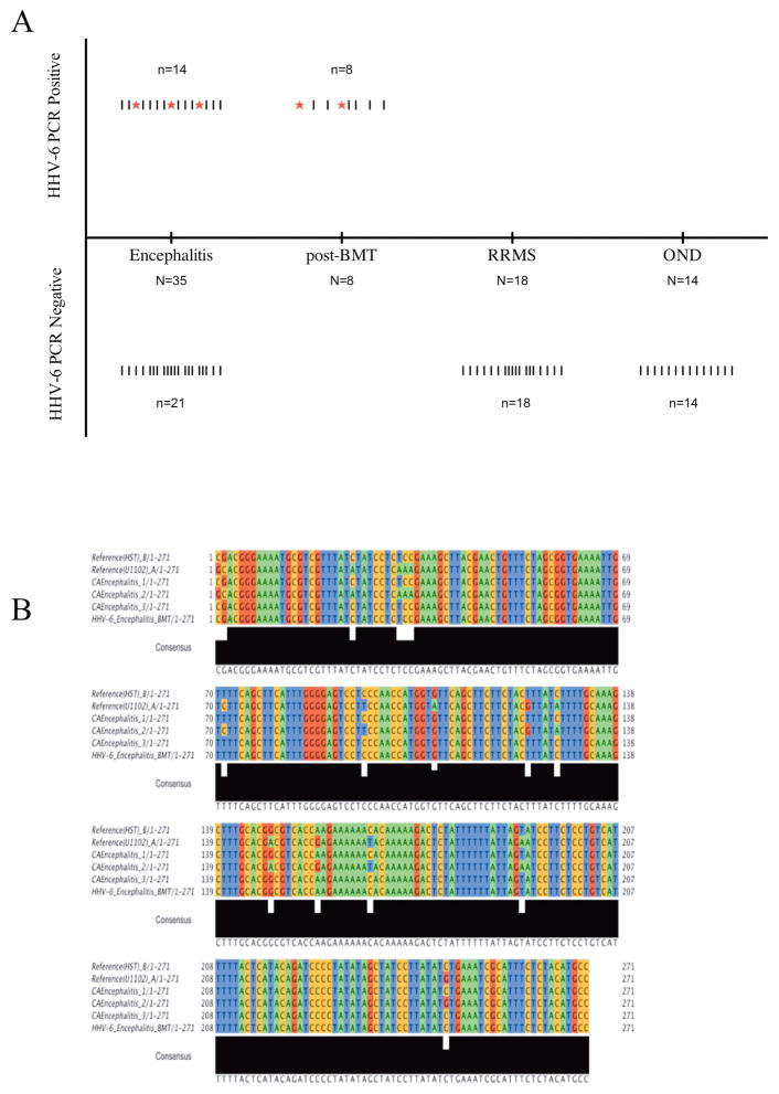
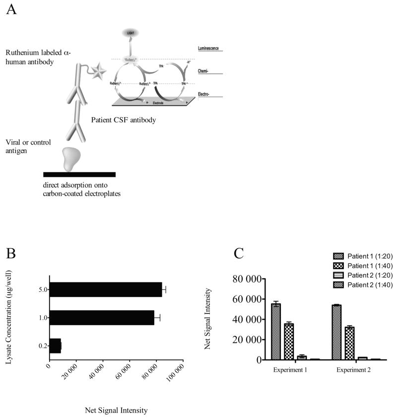
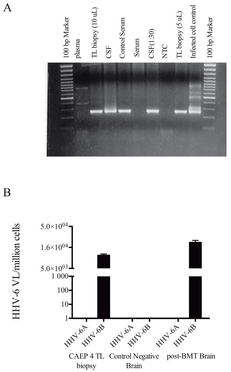
Methods: To investigate whether human herpesvirus-6 (HHV-6) is a causative agent of encephalitis, we examined for evidence of virus infection by determining the presence of viral sequence using polymerase chain reaction and assessed HHV-6 antibody reactivity in the cerebrospinal fluid of encephalitis patients with unknown cause. In a cohort study, we compared virus-specific antibody levels in cerebrospinal fluid samples of patients with encephalitis, relapsing-remitting multiple sclerosis, and other neurological diseases.

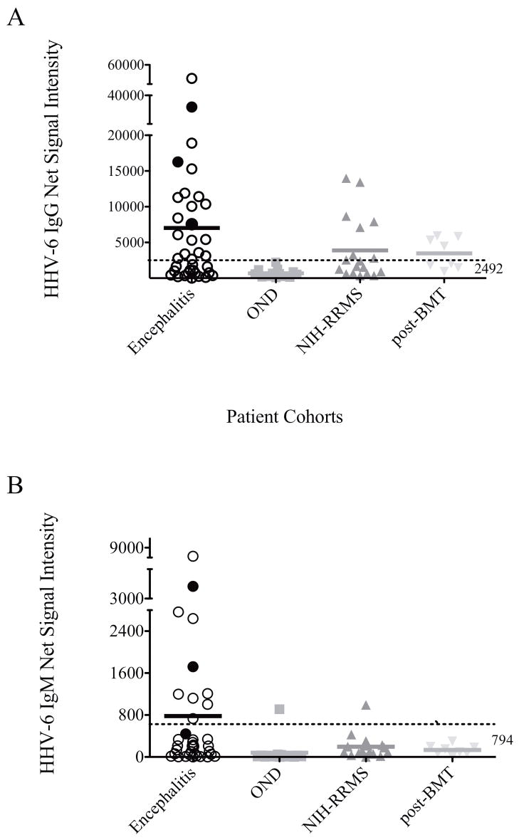
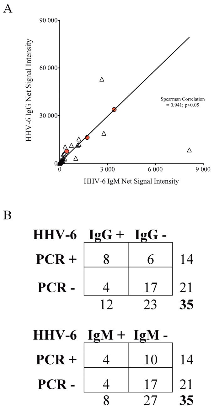
Results: Our results demonstrated increased levels of HHV-6 IgG, as well as IgM levels, in a subset of encephalitis patients compared with other neurological diseases. Moreover, cell-free viral DNA that is indicative of active infection was detected in 40% (14/35) of encephalitis patients, whereas no amplifiable viral sequence was found in either relapsing-remitting MS or other neurological diseases patients. In addition, a significant correlation between polymerase chain reaction detection and anti-HHV-6 antibody response was also demonstrated.

Interpretation: Collectively, these results suggested HHV-6 as a possible pathogen in a subset of encephalitis cases.


Figures

Figure 1
Figure 1. PCR amplication of HHV-6 sequence from CSF of encephalitis patients
A) Results of HHV-6 U57 nested PCR from various neurologic cohorts. “|” denotes each patient sample tested. “*” denotes samples that were both positive by primary and nested PCR amplication. B) Alignment of HHV-6 U12 ORF amplified by primary PCR from CSF of encephalitis patients. PCR amplicons from patients CEP1, 2 and 3 were aligned against HHV-6 A (U1102) and -6B (HST) RefSeq sequences from Genbank. CEP1 and 3 demonstrated homology to the HST strain of HHV-6 while CEP 2 showed homology to the U1102 strain.
Figure 2
Figure 2. Validation of HHV-6 Antibody ECL assay
A) Schematic diagram representing HHV-6 antibody ECL assay. Briefly, lysate from either HHV-6 infected cells or control non-infected parental cell was deposited onto the carbon-coated electroplates. Patient CSF at 1:20 or 1:40 dilution was then added and allowed to incubate for 1 hour. For detection, a ruthenium labeled anti-human IgG was added. Electrical charged is applied to the electroplate for excitation of bound ruthenium molecules. B) Validation of the ECL assay using a specific monoclonal antibody against HHV-6 P41 protein. Ruthenium labeled anti-P41 antibody was added at 1ug/mL concentration to increasing amount of lysate as represented and reactivity was measured. Net signal intensity was calculated by [Value of HHV-6 lysate - value of SupT1 lysate]. C) Representative replicate experiments demonstrated high reproducibility. HHV-6 IgG in CSF samples from two patients were tested in separate experiments at 1:20 and 1:40 dilutions. Comparable results were obtained in these independent experiments from the same samples.
Figure 3
Figure 3. Elevated HHV-6 IgG and IgM in CSF of patients with encephalitis
A) Antibody against HHV-6 in CSF of patient with encephalitis was significantly elevated compared with patients with other neurological diseases (OND). Elevated IgG was observed in NIH-RRMS patients and post-BMT recipients. The “normal range” was determined to be 2942 as defined by 3 standard deviations above the mean of OND patients. Antibody levels of CEP1,2, and 3 who demonstrated amplifiable HHV-6 sequence by primary PCR are indicated by filled circles. B) HHV-6 IgM in CSF of patient with encephalitis was specifically elevated compared with other neurological diseases cohorts. The “normal range” was determined to be 794 as defined by 3 standard deviations above the mean of OND patients. Antibody levels of CEP1,2, and 3 who demonstrated amplifiable HHV-6 sequence by primary PCR are indicated by filled circles.
Figure 4
Figure 4. Correlation of HHV-6 antibody titers and PCR detection
A) Significant correlation was demonstrated between levels of HHV-6 IgG and IgM in patients with encephalitis (spearman correlation=0.941; p<0.05). Filled circles represent HHV-6 IgG and IgM of CEP1,2, and 3. B) Statistically significant correlation was observed between HHV-6 PCR detection and HHV-6 IgG (Fischer’s exact test p=0.03) but not with HHV-6 IgM (Fischer’s exact test p=0.69).
Figure 5
Figure 5. HHV-6 detection in temporal lobe biopsy of a patient with unknown origin of encephalitis
A) HHV-6 U57 nested PCR amplification of plasma, temporal lobe biopsy, CSF, and serum samples from a patient with unknown origin of encephalitis. The results showed specific amplification of HHV-6 sequence in the CNS compartments and not in the periphery. B) HHV-6 virus load in the temporal lobe biopsy was measured by HHV-6 strain specific quantitative TaqMan real-time PCR. As shown, temporal lobe biopsy from CEP4 demonstrated 1x10⁴ copies of HHV-6B/million cells while no appreciable HHV-6A virus was detected. As controls, a specimen from a non-HHV-6 related tumor biopsy and a specimen from a post-BMT patient with neurologic complications were used as negative and positive controls, respectively.

Comment in

References

    1. Glaser CA, Honarmand S, Anderson LJ, et al. Beyond viruses: clinical profiles and etiologies associated with encephalitis. Clin Infect Dis. 2006;43:1565–1577. - PubMed
    1. Kennedy PG. Viral encephalitis: causes, differential diagnosis, and management. J Neurol Neurosurg Psychiatry. 2004;75(Suppl 1):i10–15. - PMC - PubMed
    1. Glaser CA, Gilliam S, Schnurr D, et al. In search of encephalitis etiologies: diagnostic challenges in the California Encephalitis Project, 1998–2000. Clin Infect Dis. 2003;36:731–742. - PubMed
    1. Lusso P. HHV-6 and the immune system: mechanisms of immunomodulation and viral escape. J Clin Virol. 2006;37(Suppl 1):S4–10. - PubMed
    1. Froberg MK. Review: CMV escapes! Ann Clin Lab Sci. 2004;34:123–130. - PubMed

Publication types

MeSH terms

LinkOut - more resources