

All-atom model for stabilization of alpha-helical structure in peptides by hydrocarbon staples
- PMID: 19334772
- PMCID: PMC2735086
- DOI: 10.1021/ja805037p
All-atom model for stabilization of alpha-helical structure in peptides by hydrocarbon staples
Abstract
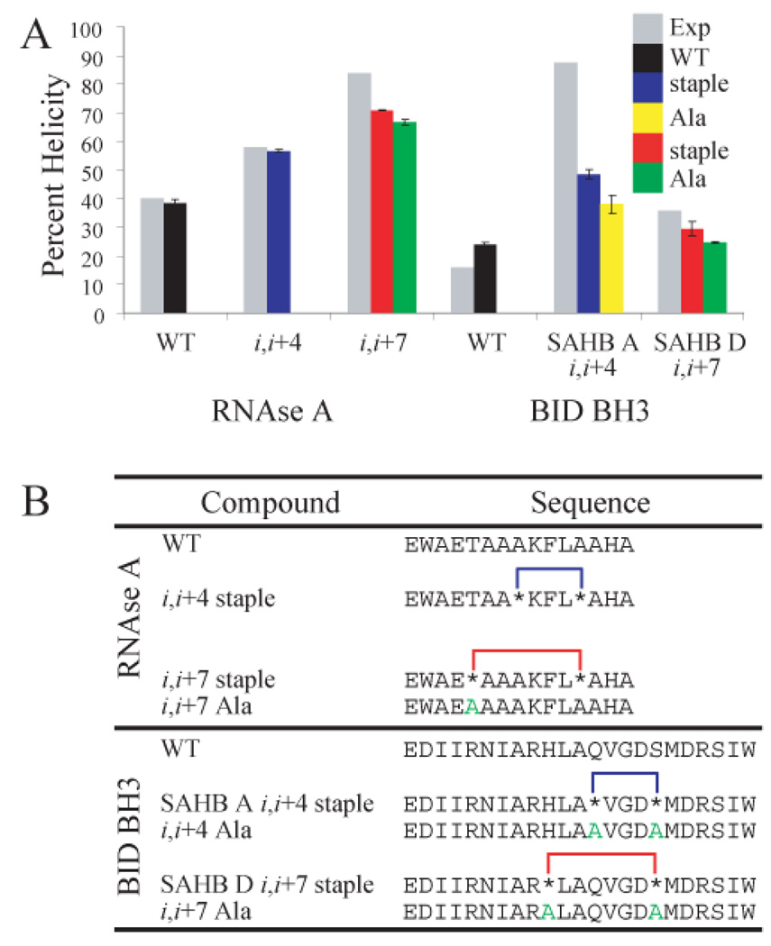
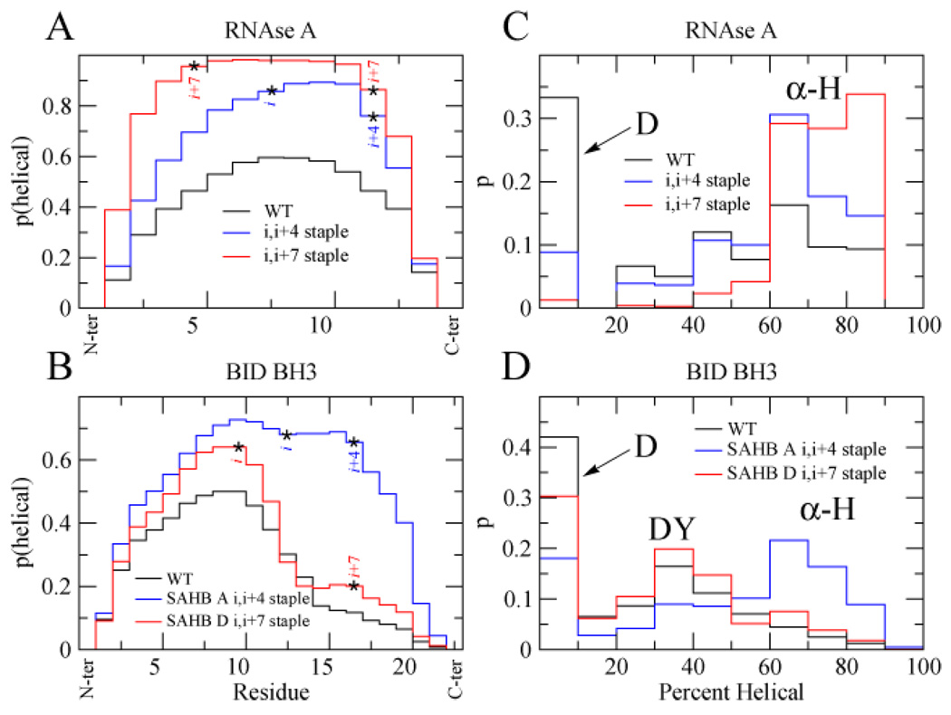
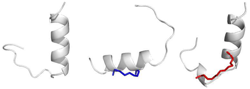
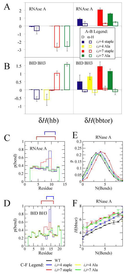
Recent work has shown that the incorporation of an all-hydrocarbon "staple" into peptides can greatly increase their alpha-helix propensity, leading to an improvement in pharmaceutical properties such as proteolytic stability, receptor affinity, and cell permeability. Stapled peptides thus show promise as a new class of drugs capable of accessing intractable targets such as those that engage in intracellular protein-protein interactions. The extent of alpha-helix stabilization provided by stapling has proven to be substantially context dependent, requiring cumbersome screening to identify the optimal site for staple incorporation. In certain cases, a staple encompassing one turn of the helix (attached at residues i and i+4) furnishes greater helix stabilization than one encompassing two turns (i,i+7 staple), which runs counter to expectation based on polymer theory. These findings highlight the need for a more thorough understanding of the forces that underlie helix stabilization by hydrocarbon staples. Here we report all-atom Monte Carlo folding simulations comparing unmodified peptides derived from RNase A and BID BH3 with various i,i+4 and i,i+7 stapled versions thereof. The results of these simulations were found to be in quantitative agreement with experimentally determined helix propensities. We also discovered that staples can stabilize quasi-stable decoy conformations, and that the removal of these states plays a major role in determining the helix stability of stapled peptides. Finally, we critically investigate why our method works, exposing the underlying physical forces that stabilize stapled peptides.
Figures
References
-
- Ghalit N, Poot AJ, Furstner A, Rijkers DTS, Liskamp RM. J. Org. Lett. 2005;7:2961–2964. - PubMed
-
- Ghalit N, Rijkers DTS, Kemmink J, Versluis C, Liskamp RM. J. Chem. Commun. 2005:192–194. - PubMed
-
- Angell Y, Burgess K. J. Org. Chem. 2005;70:9595–9598. - PubMed
-
- Miller SJ, Blackwell HE, Grubbs RH. J. Am. Chem. Soc. 1996;118:9606–9614.
-
- Fink BE, Kym PR, Katzenellenbogen JA. J. Am. Chem. Soc. 1998;120:4334–4344.
Publication types
MeSH terms
Substances
Grants and funding
LinkOut - more resources
Full Text Sources
Other Literature Sources
