. 2009 Mar 31:10:24.
doi: 10.1186/1471-2121-10-24.

Transient in utero disruption of cystic fibrosis transmembrane conductance regulator causes phenotypic changes in alveolar type II cells in adult rats

Affiliations

Transient in utero disruption of cystic fibrosis transmembrane conductance regulator causes phenotypic changes in alveolar type II cells in adult rats

Ashraf Gad et al. BMC Cell Biol. .

Abstract

Background: Mechanicosensory mechanisms regulate cell differentiation during lung organogenesis. We have previously demonstrated that cystic fibrosis transmembrane conductance regulator (CFTR) was integral to stretch-induced growth and development and that transient expression of antisense-CFTR (ASCFTR) had negative effects on lung structure and function. In this study, we examined adult alveolar type II (ATII) cell phenotype after transient knock down of CFTR by adenovirus-directed in utero expression of ASCFTR in the fetal lung.

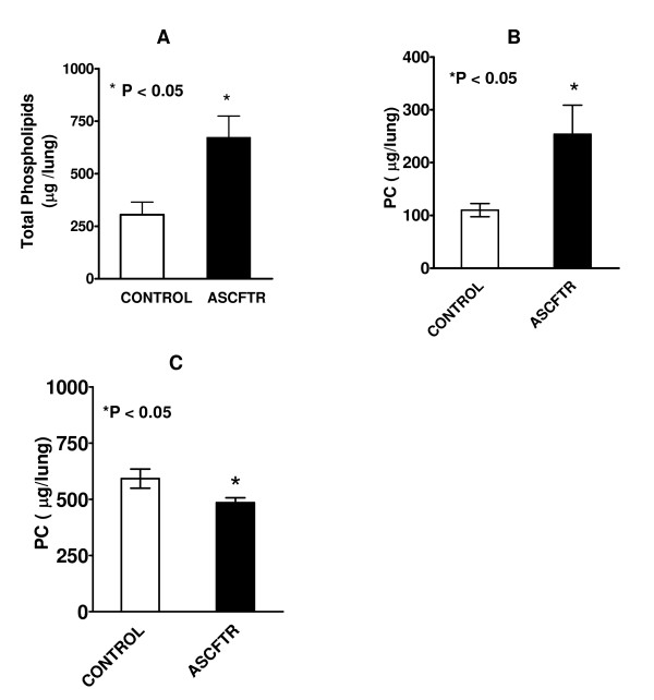
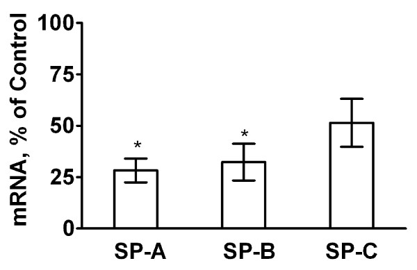
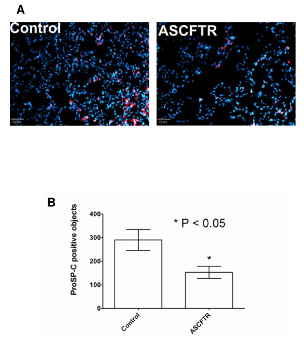
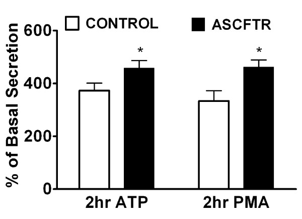
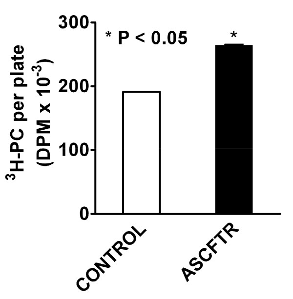
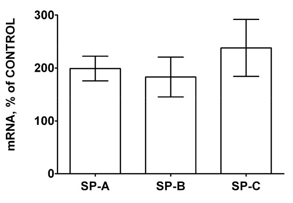
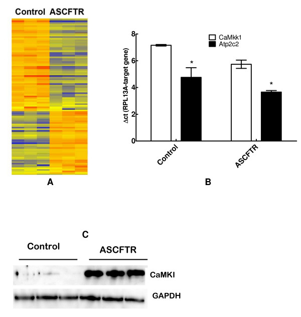
Results: In comparison to (reporter gene-treated) Controls, ASCFTR-treated adult rat lungs showed elevated phosphatidylcholine (PC) levels in the large but not in the small aggregates of alveolar surfactant. The lung mRNA levels for SP-A and SP-B were lower in the ASCFTR rats. The basal PC secretion in ATII cells was similar in the two groups. However, compared to Control ATII cells, the cells in ASCFTR group showed higher PC secretion with ATP or phorbol myristate acetate. The cell PC pool was also larger in the ASCFTR group. Thus, the increased surfactant secretion in ATII cells could cause higher PC levels in large aggregates of surfactant. In freshly isolated ATII cells, the expression of surfactant proteins was unchanged, suggesting that the lungs of ASCFTR rats contained fewer ATII cells. Gene array analysis of RNA of freshly isolated ATII cells from these lungs showed altered expression of several genes including elevated expression of two calcium-related genes, Ca2+-ATPase and calcium-calmodulin kinase kinase1 (CaMkk1), which was confirmed by real-time PCR. Western blot analysis showed increased expression of calmodulin kinase I, which is activated following phosphorylation by CaMkk1. Although increased expression of calcium regulating genes would argue in favor of Ca2+-dependent mechanisms increasing surfactant secretion, we cannot exclude contribution of alternate mechanisms because of other phenotypic changes in ATII cells of the ASCFTR group.

Conclusion: Developmental changes due to transient disruption of CFTR in fetal lung reflect in altered ATII cell phenotype in the adult life.


Figures

Figure 1
Figure 1
In utero treatment with ASCFTR causes structural changes in the lung of adult rats. Lung sections from 3 months old rats were stained with hematoxylin and eosin and viewed by light microscopy. Frequent areas showing airway thickening and multilayered epithelia (filled arrows) were observed in the ASCFTR rat lungs (B and D), while the control lungs showed normal architecture (A and C). Some alveolar areas (open arrows) with cellular infiltration were also observed in the ASCFTR rats. Original magnification, A and B ×100; C and D ×200
Figure 2
Figure 2
Phospholipid and Phosphatidylcholine in large and small aggregates of alveolar surfactant. Bronchoalveolar lavage fluid from control and ASCFTR animals was centrifuged to obtain the large and small aggregate fractions of alveolar surfactant. Lipid extracts were evaluated for total phospholipids (A) and phosphatidylcholine (PC) (B). Results are mean ± SE of 7 animals in each group and are expressed as total μg lipid in lung. Total phospholipids and PC were higher in the ASCFTR rats. * P < 0.05. The mass of phospholipid was calculated by assuming that phospholipids phosphorous comprised 4% of the lipid mass. (C) The PC levels were lower in the small aggregate (SA) fraction in the ASCFTR rats in comparison to controls (* P < 0.05, n = 7, each).
Figure 3
Figure 3
Real-time PCR of surfactant-associated protein mRNA expression in lung. Lung tissue was stored in the RNALater® buffer for extraction of total RNA. Real-time PCR was performed for quantification of mRNA levels for SP-A, SP-B and SP-C and normalized to the house-keeping gene RPL13A. To obtain ratios of normalized mRNA levels for each protein for the ASCFTR and control lung, samples were paired with respect to the day of sacrifice. The average results of triplicate samples are paired and calculated as percent of control. Results are expressed as percent change (in comparison to the control) and are mean ± SE of 5 pairs of animals for each protein. * The difference was considered significant at ≥ 50% decrease.
Figure 4
Figure 4
Immuno-staining of lung sections for proSP-C. A. Sections of frozen lungs from Control and ASCFTR rats were probed with polyclonal antibodies to proSP-C and then with Cy3-labeled secondary antibodies (red). The cell nuclei were counter-stained with DAPI (blue). The sections were viewed with a fluorescence microscope and photographed after deconvolusion and normalization of Cy3 fluorescence intensity to the same range of pixel density for the Control and ASCFTR specimens. B. A comparison of fluorescent object count between the Control and ASCFTR specimens showed a significant decrease in proSP-C positive cells (ATII cells) in the ASCFTR group in comparison to the Control group.
Figure 5
Figure 5
Lung surfactant secretion in alveolar type II cells isolated from control and ASCFTR rats. Alveolar type II cells were isolated from lungs of paired Control and ASCFTR rats. The cells were cultured for 20–22 h with [methyl-3H]choline to label cell phosphatidylcholine (PC). Following this culture period, adherent cells were incubated for 2 h without (basal secretion) or with addition of 1 mM ATP or 80 nM phorbol 12-Myristate 13-Acetate (PMA). The basal secretion was 1.05 ± 0.19% and 0.82 ± 0.11%, n = 6, in the control and ASCFTR cells, respectively (P > 0.05). The ATP and PMA-stimulated secretion is expressed as percent of the basal secretion. * P < 0.05 in comparison to the Control cells.
Figure 6
Figure 6
Phosphatidylcholine pool in alveolar type II cells. Equilibrium labeling of cell phosphatidylcholine (PC) with [methyl-3H]choline was used to measure the PC pool in alveolar type II cells. The cells were labeled for 20–22 h, as discussed for PC secretion studies. The radioactivity in the cell lipids was quantified. Results are mean ± SE of 6 separate experiments. The PC pool in ASCFTR cells was higher in comparison to the control cells. * P < 0.05.
Figure 7
Figure 7
Surfactant protein mRNA levels in alveolar type II cells. Real-time PCR using total RNA from freshly isolated (uncultured) AT II cells was performed for quantification of mRNA levels of SP-A, SP-B and SP-C (n = 6, each). The results of triplicate analysis for each sample were paired between the control and ASCFTR samples and expressed as percentage of control. The mRNA levels (normalized to the house keeping gene RPL13A) for all three proteins were similar in the two groups. The results were considered significant at ≥ 200% (≥ 2-fold) of the Control.
Figure 8
Figure 8
Gene array analysis of ATII cell RNA. Gene expression in adult type II cells isolated from animals that were infected in utero with adenovirus containing EGFP (Control) or ASCFTR constructs. A. Heat map for 101 genes in three separate preparations of type II cells demonstrating altered expression in the ASCFTR group in comparison to the Control group. B. Results of real-time PCR analysis of RNA from three separate preparations of freshly isolated type II cells showing enhanced expression of Ca2+-ATPase and calcium-dependent calmodulin kinase kinase1. Results (mean ± SE, n = 3) are expressed as change in threshold cycle for the target gene in comparison to the house keeping gene, RPL13A. A smaller ΔCt value indicates higher expression. * P < 0.05 in comparison to corresponding Control group. C. Western blot analysis showing elevated expression of calmodulin kinase I (upper panel) relative to house-keeping protein GAPDH (lower panel). The results of real-time PCR and Western blot analysis support the findings of Gene array analysis.

Similar articles

Cited by

References

    1. Kaplan F. Molecular determinants of fetal lung organogenesis. Mol Genet Metab. 2000;71:321–41. doi: 10.1006/mgme.2000.3040. - DOI - PubMed
    1. Edwards YS. Stretch stimulation: its effects on alveolar type II cell function in the lung. Comp Biochem Physiol A Mol Integr Physiol. 2001;129:245–60. doi: 10.1016/S1095-6433(01)00321-X. - DOI - PubMed
    1. Foster CD, Varghese LS, Skalina RB, Gonzales LW, Guttentag SH. In vitro transdifferentiation of human fetal type II cells toward a type I-like cell. Pediatr Res. 2007;61:404–9. doi: 10.1203/pdr.0b013e3180332c6d. - DOI - PMC - PubMed
    1. Larson JE, Cohen JC. Developmental paradigm for early features of cystic fibrosis. Pediatr Pulmonol. 2005;40:371–7. doi: 10.1002/ppul.20169. - DOI - PubMed
    1. Jiang X, Ingbar DH, O'Grady SM. Adrenergic regulation of ion transport across adult alveolar epithelial cells: effects on Cl- channel activation and transport function in cultures with an apical air interface. J Membr Biol. 2001;181:195–204. - PubMed

Publication types

MeSH terms

Substances

LinkOut - more resources