. 2009 Jun 15;18(12):2241-56.
doi: 10.1093/hmg/ddp160. Epub 2009 Mar 31.

Inverted duplications on acentric markers: mechanism of formation

Affiliations

Inverted duplications on acentric markers: mechanism of formation

Andrea E Murmann et al. Hum Mol Genet. .

Abstract

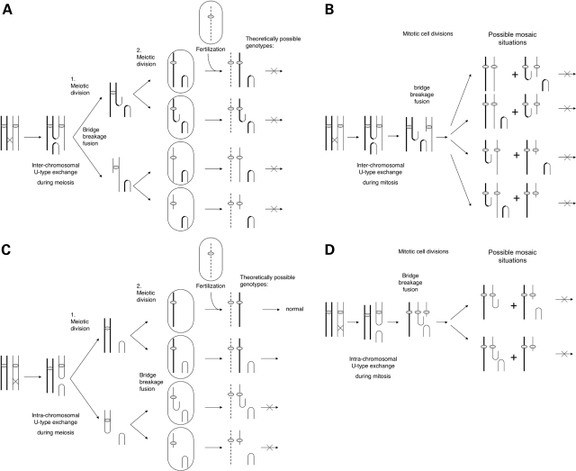
Acentric inverted duplication (inv dup) markers, the largest group of chromosomal abnormalities with neocentromere formation, are found in patients both with idiopathic mental retardation and with cancer. The mechanism of their formation has been investigated by analyzing the breakpoints and the genotypes of 12 inv dup marker cases (three trisomic, six tetrasomic, two polysomic and one X chromosome derived marker) using a combination of fluorescence in situ hybridization, quantitative SNP array and microsatellite analysis. Inv dup markers were found to form either symmetrically with one breakpoint or asymmetrically with two distinct breakpoints. Genotype analyses revealed that all inv dup markers formed from one single chromatid end. This observation is incompatible with the previously suggested model by which the acentric inv dup markers form through inter-chromosomal U-type exchange. On the basis of the identification of DNA sequence motifs with inverted homologies within all observed breakpoint regions, a new general mechanism is proposed for the acentric inv dup marker formation: following a double-strand break an acentric fragment forms, during either meiosis or mitosis. The open DNA end of the acentric fragment is stabilized by the formation of an intra-chromosomal loop promoted by the presence of sequences with inverted homologies. Likely coinciding with the neocentromere formation, this stabilized fragment is duplicated during an early mitotic event, insuring the marker's survival during cell division and its presence in all cells.


Figures

Figure 1.
Figure 1.
Determination of the breakpoint region for the acentric inv dup marker-5 that is located at 8q23.3 between 115.6 and 116.0 Mb. (AC) FISH analysis to determine the chromosomal origin of the marker and to narrow down the breakpoint region. Open arrowheads mark the two normal chromosomes 8, solid arrowheads mark the inv dup marker. (C) Alignment of clones used in the FISH analysis of the breakpoint region. The red hatched clone G248P82578F4 showed a reduced FISH signal on the marker. The black dotted rectangle highlights the FISH determined breakpoint region. (D) Log 2 ratio plot on chr8: 115.6–116 kb. The black dotted rectangle highlights the array-determined breakpoint. The blue line indicates the calculated probability of the breakpoint to be 11% between SNP_A_1994739 and SNP_A_1824502 (chr8:115768912–115771855) and to be 77% between SNP_A_1824502 and SNP_A_1994742 (chr8:115771856–115827888).
Figure 2.
Figure 2.
Asymmetric marker-12. (A) FISH analysis of the asymmetric marker-12. Clone G248P3850B10 (green) hybridizes with two signals on the marker thus marking its duplicated region, RP11–268L7 (red) hybridized with a single signal, marking the single copy region of the marker. (B) Log 2 ratio plot showed a stepwise amplification on chromosome X. One copy was presented between chrX:1-68323173, two copies were presented between chrX: 68399526–99768351 and three copies presented between X:99854723 and Xq-telomere. Note, that the baseline for chromosome X was slightly shifted because a mix of male and female normal controls was used.
Figure 3.
Figure 3.
Determination of the breakpoint region for the trisomic acentric inv dup marker-6 and detection of an additional deletion on the der(9). (A) The ideogram of this case contains one normal chromosome 9, one deleted chromosome 9 [del (9)] and the symmetrical inv dup marker. The red stippled line marks the breakpoint of the inv dup marker, the blue-circled region points out the additional deleted part that is not present on the marker or the del(9). (B) Log 2 ratio plot on chromosome 9 shows an amplification on 9p with an additional small deletion between 32.5 and 32.8 Mb. (C and D) FISH analysis of the symmetric marker-6. (C) The partial metaphase shows the symmetrical inv dup marker (solid arrowhead), one del(9) (arrow) and one normal chromosome 9 (open arrowhead). (C) Clone G248P81068E7 (green) hybridized on the deleted part of region and is only present on the normal chromosome. G248P85568A3 (red) hybridized to the region that was present on the marker in two copies but not on the del(9). The 9p-telomeric and 9q-telomeric FISH-probes hybridize to the respective telomeres (aqua). (D) Alignment of clones used in the FISH analysis of the breakpoint regions on chromosome 9 between 34.45 and 34.9 Mb. The hatched clones G248P85622H1 showed a reduced signal on the marker, G248P81915B7 showed a reduced signal on the del (9). (E) The breakpoint regions determined by FISH and array overlap.
Figure 4.
Figure 4.
SNP-ratio analysis of chromosomal regions containing acentric inv dup markers -10 (A and B), -4 (CE), -12 (H and I), -2 (F) and -3 (G). (A, C, H) The ideograms indicate how many copies are present in the genome, the horizontal lines mark the positions of the breakpoints, the brackets span the regions in which the SNPs ratios were calculated (B, D, I, F, G). The ratios were plotted and displayed using the formula (ab)/(a+b). (E) Ratio analysis of the tetrasomic region of the acentric marker-4 was displayed according to their actual chromosomal position (chr8:0–6.8 Mb). The positions of 2:2 and 3:1 ratios were marked above the plot to better visualize the random distribution of both ratios along chr8 indicate the presence of three different haplotypes. Comparison of theoretical expected ratios from Supplementary Material, Fig. S1 with the observed ratios allowed conclusions regarding the various inv dup marker formations.
Figure 5.
Figure 5.
Alignments of selected sequence motifs with inverted homologies found in the breakpoint regions of acentric inv dup markers. (A and B) If two sequences from two different chromosomal locations were aligned the inverted homologies were represented by a line from the top left corner, to the bottom right corner. (CG) If a single sequence was aligned with itself, one line (from the bottom left to the top right) represented the sequence alignment to itself. A line from the top left to the bottom right represented its palindromic nature. Images of the alignments of the sequences were used as displayed by the BLAST2 program. The degree of homology was determined with the programs BLAT and Blast2. (A and B) Two sequences with high inverted homologies were found within the two breakpoints of the asymmetric inv dup markers -4 (A) and -12 (B). (C–G) Sequences with inverted homologies were found in the single breakpoints of the symmetric inv dup markers -1, -3, -5, -10. (C and D) Sequences with small spacer, (E–G) Sequences without spacers. (A) Alignment of two LCRs (no.23429 at chr8:6909899–6919457 and chr8:12608622–12617968) present within the two breakpoints of the asymmetric marker-4 with opposite orientation and a homology above 89%. (B) Two LINE elements (chrX:68386150–68386934 and chrX:99799609–99800394) were present within the two breakpoints of the asymmetric marker-12 with inverted homology of 73%. (C) Two inverted LINE sequences (within chr8:115812418–115817329, 77% homologous) were present within the single breakpoint of marker-5 and were separated by a spacer of ∼3 kb. (D) Palindromic SINE sequences separated by a short spacer were present on marker-10 (within chr13:65585050–65585535, 81% homologous). (E) Palindromic SINE sequences on marker-1 (chr3:183031970–183032532) is 79% homologous to its inverted complement without spacer. (F) Palindromic HSCA no. 5793340 with an AT rich center on chr9:13130211–13130484 is located on marker-3, (79–100% homologous to its inverted complement). (G) Palindromic AT rich region on chr13:65603716–65604209 is located on marker-10 with 82% homology to its inverted complement. Scale markers are shown as horizontal bars; fat bars: 2000 bp, thin bars: 200 bp.
Figure 6.
Figure 6.
The previously postulated U-type exchange mechanisms suggested how the acentric inv dup markers could form in patients with idiopathic mental retardation. Most theoretically possible genotype situations after fertilization have not been experimentally observed (marked with crosses on arrows). (A and B) The U-type exchange could either occur between two chromatid of two different chromosomes (inter-chromosomal). (C and D) It could also occur between two chromatids of the same chromosome (intra-chromosomal). During meiosis crossing over leads to the formation of one acentric inv dup marker with two arms and one dicentric derivative. After meiosis I and the meiosis II division, respectively, the inter- and intra-chromosomal dicentrics break when both their centromeres are pulled apart resulting in a deleted derivative and an inv dup del derivative. (B and D) U-type exchange during mitosis also results in theoretical genotype situations, which were never observed in this study.
Figure 7.
Figure 7.
Proposed new mechanism for the formation of inv dup marker involving from a single chromatid end. (A) After a double-strand break a chromatid end breaks off. One DNA strand of this acentric fragment is exposed by 5′ to 3′ degradation. Intra-strand pairing might be facilitated at this position due to close proximity of sequences with inverted homology and subsequently a hairpin molecule is generated. After neocentromere formation and replication, the fragment is amplified to a ‘large palindrome’: the acentric inv dup marker. Light gray ovals are the centromeres, the filled ovals are neocentromeres. The hairpin molecule prior to replication could pair with the deleted chromosome (resulting into trisomy) or with one normal chromosome (resulting into tetrasomy). (B) The break occurs prior to first meiosis I division and leads to all possible and experimentally observed situations. (C) The break occurs during the meiosis II division could also lead to all experimentally observed situations. (D) If the break occurs during a mitotic event, mosaicism for trisomic cases would result, or tetrasomic mixed with a deleted karyotype, both of which have not been observed experimentally in cases with idiopathic mental retardation.

References

    1. Italiano A., Attias R., Aurias A., Perot G., Burel-Vandenbos F., Otto J., Venissac N., Pedeutour F. Molecular cytogenetic characterization of a metastatic lung sarcomatoid carcinoma: 9p23 neocentromere and 9p23–p24 amplification including JAK2 and JMJD2C. Cancer Genet. Cytogenet. 2006;167:122–130. - PubMed
    1. Abeliovich D., Yehuda O., Ben-Neriah S., Kapelushnik Y., Ben-Yehuda D. dup(10q) lacking alpha-satellite DNA in bone marrow cells of a patient with acute myeloid leukemia. Cancer Genet. Cytogenet. 1996;89:1–6. - PubMed
    1. Sirvent N., Forus A., Lescaut W., Burel F., Benzaken S., Chazal M., Bourgeon A., Vermeesch J.R., Myklebost O., Turc-Carel C., et al. Characterization of centromere alterations in liposarcomas. Genes Chrom. Cancer. 2000;29:117–129. - PubMed
    1. Gisselsson D., Hoglund M., Mertens F., Mitelman F., Mandahl N. Chromosomal organization of amplified chromosome 12 sequences in mesenchymal tumors detected by fluorescence in situ hybridization. Genes Chrom. Cancer. 1998;23:203–212. - PubMed
    1. Marshall O.J., Chueh A.C., Wong L.H., Choo K.H. Neocentromeres: new insights into centromere structure, disease development, and karyotype evolution. Am. J. Hum. Genet. 2008;82:261–282. - PMC - PubMed

Publication types

Substances