. 2009 Apr 15;69(8):3529-36.
doi: 10.1158/0008-5472.CAN-09-0067. Epub 2009 Mar 31.

Guanylyl cyclase C prevents colon cancer metastasis by regulating tumor epithelial cell matrix metalloproteinase-9

Affiliations

Guanylyl cyclase C prevents colon cancer metastasis by regulating tumor epithelial cell matrix metalloproteinase-9

Wilhelm J Lubbe et al. Cancer Res. .

Abstract

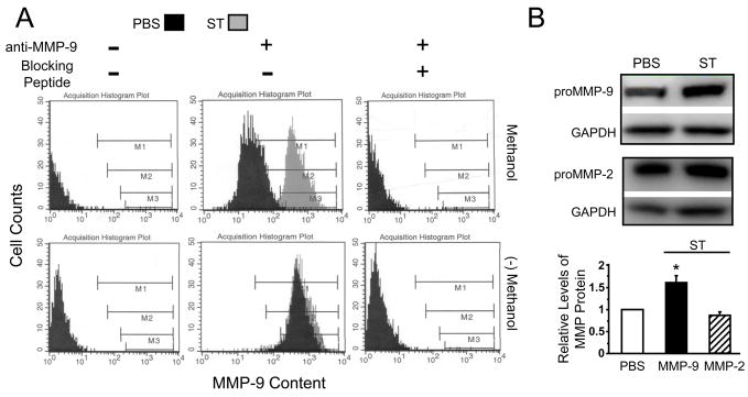
Matrix metalloproteinase-9 (MMP-9) produced by colorectal cancer cells is a critical determinant of metastatic disease progression and an attractive target for antimetastatic strategies to reduce colon cancer mortality. Cellular signaling by cyclic GMP (cGMP) regulates MMP-9 dynamics in various cell systems, and the bacterial enterotoxin receptor guanylyl cyclase C (GCC), the principle source of cGMP in colonocytes, which is overexpressed in colorectal cancers, inhibits tumor initiation and progression in the intestine. Here, we show that ligand-dependent GCC signaling through cGMP induces functional remodeling of cancer cell MMP-9 reflected by a compartmental redistribution of this gelatinase, in which intracellular retention resulted in reciprocal extracellular depletion. Functional remodeling of MMP-9 by GCC signaling reduced the ability of colon cancer cells to degrade matrix components, organize the actin cytoskeleton to form locomotory organelles and spread, and hematogenously seed distant organs. Of significance, GCC effects on cancer cell MMP-9 prevented establishment of metastatic colonies by colorectal cancer cells in the mouse peritoneum in vivo. Because endogenous hormones for GCC are uniformly deficient in intestinal tumors, reactivation of dormant GCC signaling with exogenous administration of GCC agonists may represent a specific intervention to target MMP-9 functions in colon cancer cells. The notion that GCC-mediated regulation of cancer cell MMP-9 disrupts metastasis, in turn, underscores the unexplored utility of GCC hormone replacement therapy in the chemoprevention of colorectal cancer progression.


Figures

Figure 1
Figure 1
GCC signaling depletes extracellular MMP-9 proteolytic activity. A, T84 and Caco-2 colon carcinoma cells expressing GCC (28) were treated for 24 h in serum-free DMEM/F12 media with PBS, 8-pCPT-cGMP (1 mM), 8-br-cGMP (5 mM) or ST (1 μM ST). Then, cell conditioned media were subjected to immunoblotting. B, Gelatin zymography of conditioned media from T84 cells treated as in A. Immunoblots (A, upper panel) and zymograms (B, upper panel) show representative experiments repeated ≥3 times. Bar graphs (lower panels) reflect quantification of bands in immunoblots (A) or zymograms (B) by densitometry, and represent percentages of respective vehicle (PBS)-treated control cells. *, P < 0.05; **, P < 0.01; ***, P < 0.005 versus respective control. C, Levels of MMP-9 mRNA (per μg RNA) by RT-PCR, relative to the PBS control.
Figure 2
Figure 2
GCC signaling induces intracellular MMP-9 retention. T84 cells were treated (24 h in serum-free DMEM/F12) with PBS or ST (1 μM) and subjected to flow cytometry (A) or immunoblot analysis (B). In A, intracellular MMP-9 protein was analyzed in cells permeabilized with methanol, while membrane-bound MMP-9 was quantified employing non-permeabilized cells. Histograms of MMP-9-stained cells are representative of three independent experiments. In B, upper panels show a representative experiment, while the lower panel reflects mean ± SEM of MMP-9 or MMP-2 levels in total cell lysates, normalized to the respective loading (GAPDH) and vehicle (PBS) controls, from three independent experiments as quantified by densitometry. *, P < 0.05 versus respective control (PBS).
Figure 3
Figure 3
GCC activation suppresses actin cytoskeleton-based locomotory organelles. A, Representative images (magnification ×63) from Differential Interference Contrast (DIC) and confocal microscopy of PBS- (upper panels) and ST-treated (lower panels) T84 cells. Cells were stained with 4′,6-diamidino-2-phenylindole (DAPI, for nuclei in blue), phalloidin (green) and anti-MMP-9 antibody (red). Yellow in merged images indicates β-actin and MMP-9 colocalization. B, Cell spreading was quantified as the ratio of cells extending membrane protrusions (filopodia, lamellipodia) versus the total cell number per microscopic field (magnification, 20×) of an inverted microscope (13). TJU, the inactive ST analog Thomas Jefferson University 1–103. GCC ligands and the inactive analog were employed at 1 μM each for 24 h. Data represent percent inhibition of cell spreading obtained with PBS-treated control. *, P < 0.05 versus control.
Figure 4
Figure 4
GCC signaling inhibits cell spreading by regulating MMP-9. A, Cell spreading of T84 cells treated for 24 h with ST (1 μM) or 8-br-cGMP (5 mM), in the presence or absence of the active form of MMP-9 (aMMP-9, 500 ng/ml). B–D, T84 cells were transduced with either MSCV-empty vector (T84-V) or an MSCV carrying MMP-9 (T84-MMP-9), and cell cultures stably expressing the engineered nucleic acid were selected in puromycin-supplemented media. B, Levels of MMP-9 mRNA (per μg RNA) by RT-PCR, relative to the empty vector control. C, Representative immunoblot of the MMP-9 pro-form (proMMP-9) in media conditioned by tumor cells. D, Effects of ST treatment (1 μM, 24 h) on the spreading of MSCV-transduced tumor cells. Cell spreading in A and D was quantified as described in Fig. 3B, and data represent percent inhibition of cell spreading obtained with the respective vehicle-treated control. *, P < 0.05; **, P < 0.01 versus respective control.
Figure 5
Figure 5
GCC signaling reduces hematogenous seeding of mouse lung. A, Representative inverted fluorescence microscope fields of MitoTracker-stained wild type T84 cells and isogenic clones stably expressing the MSCV-empty vector (T84-V) or the MSCV-MMP-9 vector (T84-MMP-9). Cells were treated in vitro (24 h in serum-free media) with PBS (upper panels) or ST (1 μM, lower panels). Bar, 200 μm. B, Tumor cell seeding of mouse lung in vivo was quantified as described in Methods. Results are percentages of respective vehicle-treated controls. Uroguanylin, 1 μM; 8-pCPT-cGMP, 1 mM; MMP-9, 500 ng/ml of purified human pro-MMP-9; BB94, a broad MMP-9 inhibitor (60 nmol/L). MMP-9 and BB94 were added to cell cultures during the last 2 h of incubations. With the exception of uroguanylin (N=2), at least 3 animals per condition were examined. *, P < 0.05; ***, P < 0.005 versus respective control.
Figure 6
Figure 6
Ligand-dependent GCC signaling suppresses peritoneal metastasis. A, Representative H&E slides of diaphragmatic rolls from mice injected with T84 cells treated in vitro (24 h, in serum-free media) with PBS or ST (1 μM). B, Metastatic tumor burden in the mouse peritoneum 2 weeks after intra-peritoneal injection of wild type T84 cells, or T84 cells stably expressing either the MSCV-empty vector (T84-V) or the MSCV-MMP-9 vector (T84-MMP-9). Cells were treated in vitro as in A and correspondent in vivo tumor formation in mouse diaphragms was quantified by RT-PCR for human GCC, a specific T84 cell marker (24, 25). Levels of GCC mRNA were calculated as 2[(blank Ct) − (sample Ct)]/μg of mouse β-actin mRNA, where Ct is the sample threshold cycle number of mice not injected (blank) or injected (sample) with cancer cells. Numbers of animals were: PBS, 13 (T84), 11 (T84-V) and 14 (T84-MMP-9); ST, 10 (T84), 12 (T84-V) and 11 (T84-MMP-9). *, P < 0.05 versus respective control. C, Proposed antimetastatic actions of GCC signaling in colorectal cancer (see Discussion for description).

References

    1. American Cancer Society Cancer facts & figures 2008. Atlanta: American Cancer Society; 2008.
    1. Sobrero A, Kerr D, Glimelius B, et al. New directions in the treatment of colorectal cancer: a look to the future. Eur J Cancer. 2000;36:559–66. - PubMed
    1. Meyerhardt JA, Mayer RJ. Systemic therapy for colorectal cancer. N Engl J Med. 2005;352:476–87. - PubMed
    1. Jemal A, Tiwari RC, Murray T, et al. Cancer statistics, 2004. CA Cancer J Clin. 2004;54:8–29. - PubMed
    1. Fidler IJ. The pathogenesis of cancer metastasis: the ‘seed and soil’ hypothesis revisited. Nat Rev Cancer. 2003;3:453–8. - PubMed

Publication types

MeSH terms