. 2009 Apr 15;69(8):3704-12.
doi: 10.1158/0008-5472.CAN-08-4344. Epub 2009 Mar 31.

Atg5 regulates phenethyl isothiocyanate-induced autophagic and apoptotic cell death in human prostate cancer cells

Affiliations

Atg5 regulates phenethyl isothiocyanate-induced autophagic and apoptotic cell death in human prostate cancer cells

Ajay Bommareddy et al. Cancer Res. .

Abstract

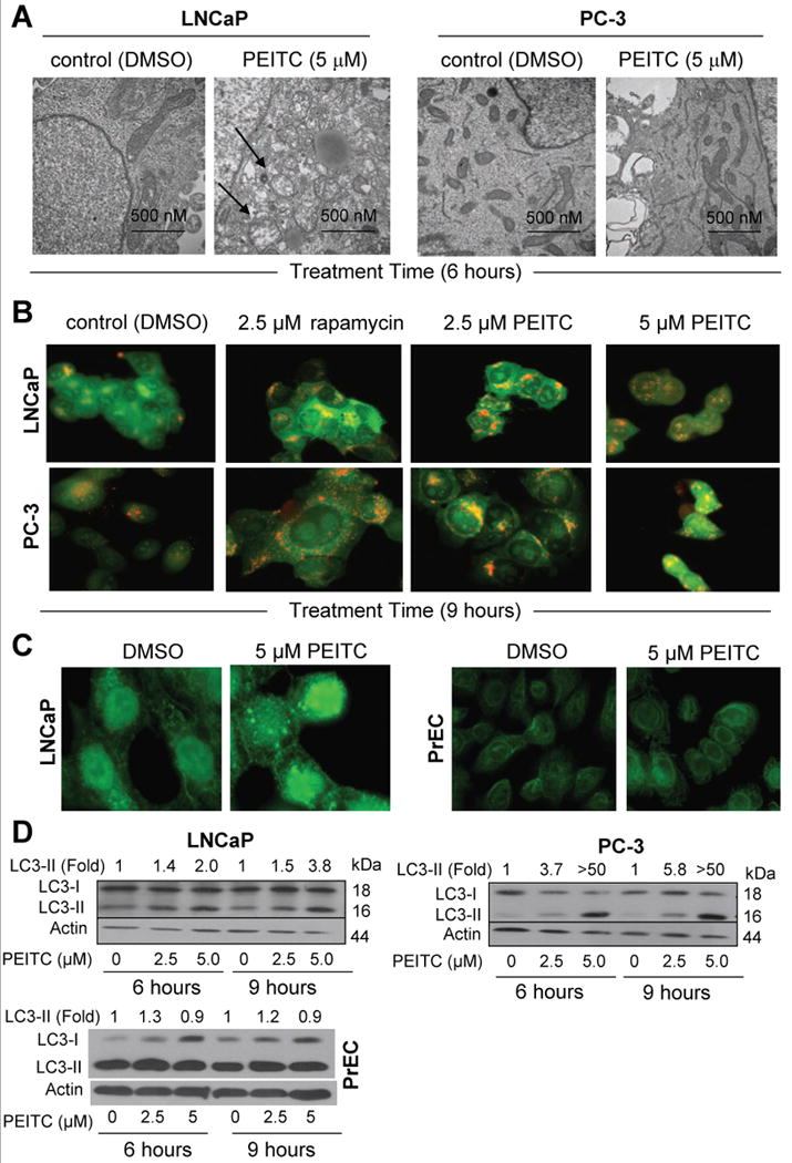
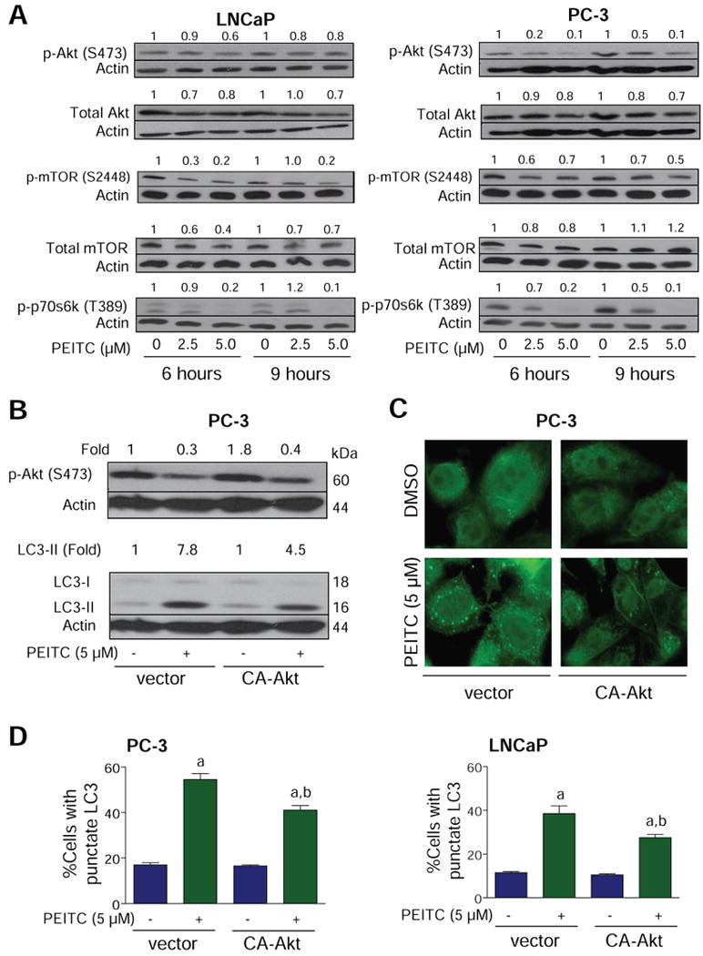
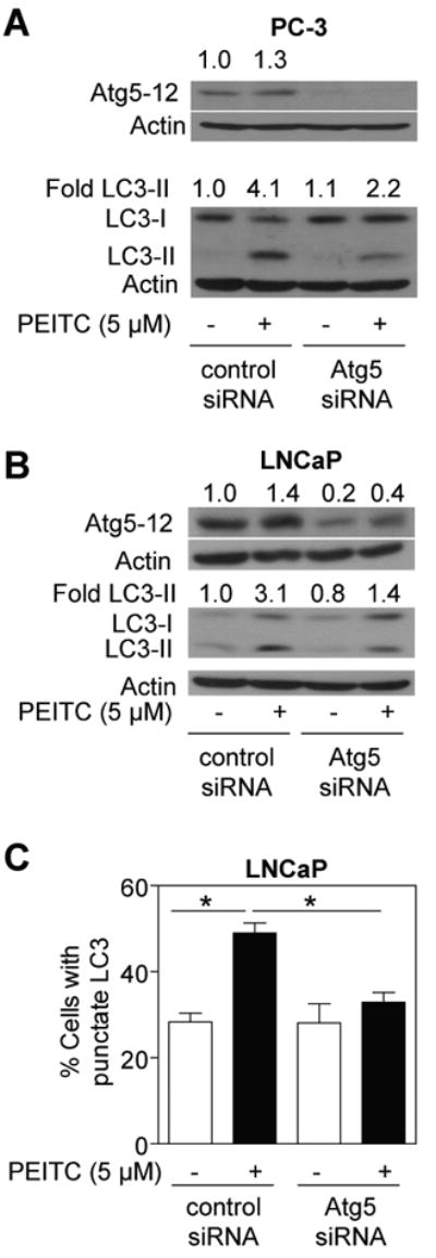
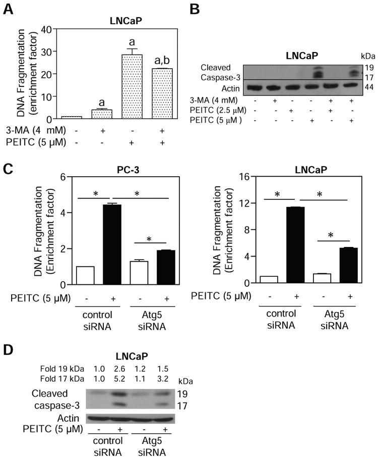
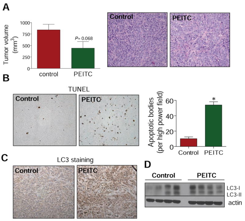
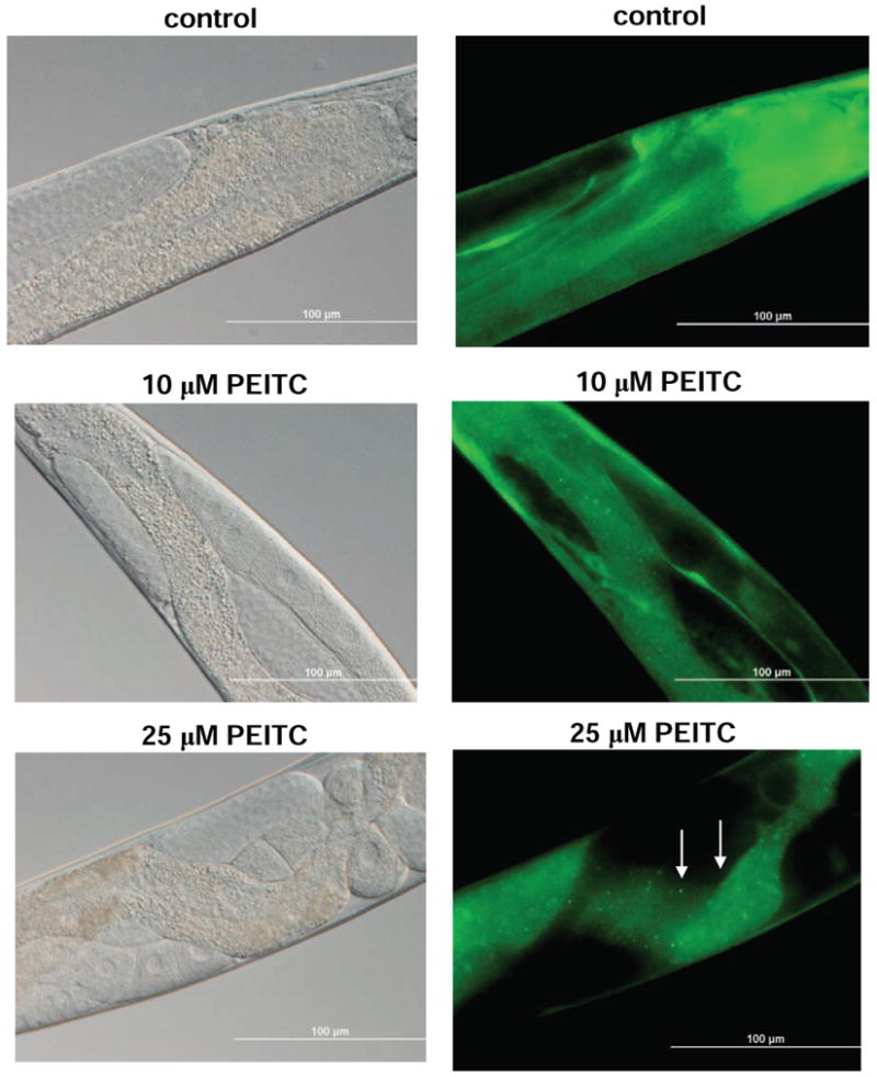
Phenethyl isothiocyanate (PEITC) is a promising cancer chemopreventive agent but the mechanism of its anticancer effect is not fully understood. We now show, for the first time, that PEITC treatment triggers Atg5-dependent autophagic and apoptotic cell death in human prostate cancer cells. Exposure of PC-3 (androgen independent, p53 null) and LNCaP (androgen responsive, wild-type p53) human prostate cancer cells to PEITC resulted in several specific features characteristic of autophagy, including appearance of membranous vacuoles, formation of acidic vesicular organelles, and cleavage and recruitment of microtubule-associated protein 1 light chain 3 (LC3) to autophagosomes. A normal human prostate epithelial cell line (PrEC) was markedly more resistant toward PEITC-mediated cleavage and recruitment of LC3 compared with prostate cancer cells. Although PEITC treatment suppressed activating phosphorylations of Akt and mammalian target of rapamycin (mTOR), which are implicated in regulation of autophagy by different stimuli, processing and recruitment of LC3 was only partially/marginally reversed by ectopic expression of constitutively active Akt or overexpression of mTOR-positive regulator Rheb. The PEITC-mediated apoptotic DNA fragmentation was significantly attenuated in the presence of a pharmacologic inhibitor of autophagy (3-methyl adenine). Transient transfection of LNCaP and PC-3 cells with Atg5-specific small interfering RNA conferred significant protection against PEITC-mediated autophagy as well as apoptotic DNA fragmentation. A xenograft model using PC-3 cells and Caenorhabditis elegans expressing a lgg-1:GFP fusion protein provided evidence for occurrence of PEITC-induced autophagy in vivo. In conclusion, the present study indicates that Atg5 plays an important role in regulation of PEITC-induced autophagic and apoptotic cell death.


Figures

Fig. 1
Fig. 1
A, representative transmission electron micrographs depicting ultrastructures of LNCaP and PC-3 cells treated with DMSO (control) or 5 μmol/L PEITC for 6 h (30,000× magnification). B, acridine orange staining in LNCaP and PC-3 cells treated with DMSO, 2.5 μmol/L rapamycin (positive control), and 2.5 or 5 μmol/L PEITC for 9 h. C, fluorescence microscopic analysis for punctate pattern of LC3 in LNCaP cells (9 h treatment) and PrEC normal prostate epithelial cells (6 h treatment) treated with DMSO or 5 μmol/L PEITC. D, immunoblotting for LC3 using lysates from LNCaP, PC-3, and PrEC cells treated for 6 or 9 h with DMSO or the indicated concentrations of PEITC. Densitometric quantitation for cleaved LC3-II relative to DMSO-treated control is shown on top of the immunoreactive band.
Fig. 2
Fig. 2
A, immunoblotting for phospho-(S473)-Akt, total Akt, phospho-(S2448)-mTOR, total mTOR, and phospho-(T389)-p70s6k using lysates from LNCaP or PC-3 cells treated with DMSO or 2.5 and 5 μmol/L PEITC for 6 or 9 h. B, immunoblotting for phospho-(S473)-Akt and LC3 using lysates from PC-3 cells transiently transfected with the empty-vector or vector encoding CA-Akt and treated with DMSO or 5 μmol/L PEITC for 9 h. Densitometric quantitation relative to DMSO-treated control is shown on top of the immunoreactive band. C, immunofluorescence microscopy for analysis of punctate pattern of LC3 localization in PC-3 cells transiently transfected with the vector encoding CA-Akt or empty-vector and treated with DMSO (control) or 5 μmol/L PEITC for 9 h. D, percentage of cells with punctate LC3 in PC-3 or LNCaP cultures transiently transfected with the empty-vector or vector encoding CA-Akt and treated with DMSO (control) or 5 μmol/L PEITC for 9 h. Columns, mean (n= 3); bars, SE. Significantly different (P<0.05) compared with acorresponding DMSO-treated control and bPEITC-treated empty-vector transfected cells by one-way ANOVA followed by Bonferroni's multiple comparison test. Each experiment was repeated twice and the results were comparable.
Fig. 3
Fig. 3
Immunoblotting for Atg5-12 and LC3 using lysates from (A) PC-3 cells and (B) LNCaP cells transiently transfected with a control non-specific siRNA or a pool of 3 Atg5-targeted siRNA and treated with DMSO or 5 μmol/L PEITC for 9 h. Densitometric quantitation relative to DMSO-treated non-specific siRNA transfected cells is shown on top of the immunoreactive band. C, percentage of cells with punctate LC3 in LNCaP cells transiently transfected with a control non-specific siRNA or Atg5-targeted siRNA and treated with DMSO (control) or 5 μmol/L PEITC for 9 h. *Significantly different (P<0.05) between the indicated groups by one-way ANOVA followed by Bonferroni's test. Each experiment was repeated twice and the results were comparable.
Fig. 4
Fig. 4
A, quantitation of cytoplasmic histone-associated DNA fragmentation in LNCaP cells following 16 h treatment with 5 μmol/L PEITC in the absence or presence of 4 mmol/L 3-MA (2 h pre-treatment). Columns, mean (n=3); bars, SE. Significantly different (P<0.05) compared with aDMSO-treated control and bPEITC alone treatment group by one-way ANOVA followed by Bonferroni's multiple comparison test. B, immunoblotting for cleaved caspase-3 using lysates from LNCaP cells treated for 16 h with 2.5 or 5 μmol/L PEITC in the absence or presence of 4 mmol/L 3-MA (2 h pre-treatment). C, cytoplasmic histone-associated DNA fragmentation in PC-3 (left panel) and LNCaP cells (right panel) transiently transfected with a control non-specific siRNA or a pool of 3 Atg5-targeted siRNA and treated with DMSO (control) or 5 μmol/L PEITC for 9 h. Columns, mean (n=3); bars, SE. *Significantly different (P<0.05) between the indicated groups by one-way ANOVA followed by Bonferroni's test. D, immunoblotting for cleaved caspase-3 using lysates from LNCaP cells transiently transfected with a control non-specific siRNA or a pool of 3 Atg5-targeted siRNA and treated with DMSO (control) or 5 μmol/L PEITC for 9 h. Each experiment was repeated at least twice and the results were comparable.
Fig. 5
Fig. 5
A, tumor volume in vehicle-treated control mice and PEITC-treated mice. Columns, mean (n= 5); bars, SE. Right panel, H&E staining in representative tumor section of a vehicle-treated control mouse and tumor section of a PEITC-treated mouse. B, visualization of TUNEL-positive apoptotic bodies in representative tumor section of a vehicle-treated control mouse and tumor section of a PEITC-treated mouse. Right panel, quantitation of TUNEL-positive apoptotic bodies in tumor sections from control mice and PEITC-treated mice. Columns, mean (n= 3); bars, SE. *Significantly different (P<0.05) compared with control by t-test. C, LC3 expression in representative tumor section of a vehicle-treated control mouse and tumor section of a PEITC-treated mouse. D, immunoblotting for LC3 using tumor supernatants from four different mice of both vehicle-treated control and PEITC-treated groups.
Fig. 6
Fig. 6
Representative bright field (left) and fluorescence (right) images showing cellular localization of lgg-1:GFP in intestinal cells of control (DMSO-treated) and PEITC-fed worms. In contrast to controls, the worms fed 10 or 25 μmol/L PEITC for 24 h exhibited redistribution of lgg-1:GFP from a diffuse cytoplasmic pattern to punctate foci representing the pre- and autophagosomal structures (marked by arrows). Images were captured at 40× objective lens magnification. Bars, 100 μm.

References

    1. Nelson WG, De Marzo AM, Isaacs WB. Prostate Cancer. N Engl J Med. 2003;349:366–81. - PubMed
    1. Verhoeven DT, Goldbohm RA, van Poppel G, Verhagen H, van den Brandt PA. Epidemiological studies on brassica vegetables and cancer risk. Cancer Epidemiol Biomarkers Prev. 1996;5:733–48. - PubMed
    1. Kolonel LN, Hankin JH, Whittemore AS, et al. Vegetables, fruits, legumes and prostate cancer: a multiethnic case-control study. Cancer Epidemiol Biomarkers Prev. 2000;9:795–804. - PubMed
    1. Hecht SS. Inhibition of carcinogenesis by isothiocyanates. Drug Metab Rev. 2000;32:395–411. - PubMed
    1. Conaway CC, Yang YM, Chung FL. Isothiocyanates as cancer chemopreventive agents: their biological activities and metabolism in rodents and humans. Curr Drug Metab. 2002;3:233–55. - PubMed

Publication types

MeSH terms