. 2009 Apr 14;106(15):6291-6.
doi: 10.1073/pnas.0900107106. Epub 2009 Mar 31.

Mutations in the beta-myosin rod cause myosin storage myopathy via multiple mechanisms

Affiliations

Mutations in the beta-myosin rod cause myosin storage myopathy via multiple mechanisms

Thomas Z Armel et al. Proc Natl Acad Sci U S A. .

Abstract

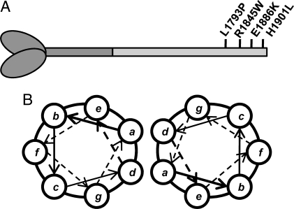
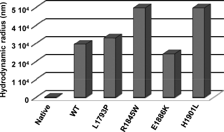
Myosin storage myopathy (MSM) is a congenital myopathy characterized by the presence of subsarcolemmal inclusions of myosin in the majority of type I muscle fibers, and has been linked to 4 mutations in the slow/cardiac muscle myosin, beta-MyHC (MYH7). Although the majority of the >230 disease causing mutations in MYH7 are located in the globular head region of the molecule, those responsible for MSM are part of a subset of MYH7 mutations that are located in the alpha-helical coiled-coil tail. Mutations in the myosin head are thought to affect the ATPase and actin-binding properties of the molecule. To date, however, there are no reports of the molecular mechanism of pathogenesis for mutations in the rod region of muscle myosins. Here, we present analysis of 4 mutations responsible for MSM: L1793P, R1845W, E1886K, and H1901L. We show that each MSM mutation has a different molecular phenotype, suggesting that there are multiple mechanisms by which MSM can be caused. These mechanisms range from thermodynamic and functional irregularities of individual proteins (L1793P), to varying defects in the assembly and stability of filaments formed from the proteins (R1845W, E1886K, and H1901L). In addition to furthering our understanding of MSM, these observations provide the first insight into how mutations affect the rod region of muscle myosins, and provide a framework for future studies of disease-causing mutations in this region of the molecule.


Conflict of interest statement

The authors declare no conflict of interest.

Figures

Fig. 1.
Fig. 1.
Location of MSM mutations in the coiled-coil rod region of β-MyHC. (A) Schematic of β-MyHC structure. The globular head region and N-terminal portion of the myosin rod which comprise heavy meromyosin are shown in dark gray. The C-terminal LMM region of the rod is shown in light gray with the relative position of MSM mutations in the distal portion of the rod marked. (B) Diagram of the relative positions of amino acids within the coiled-coil. The position of amino acids within the heptad is denoted by a–g. Three MSM mutations (R1845W, E1886K, and H1901L) are located in the outer f position, whereas 1 (L1793P) is located in the inner d position.
Fig. 2.
Fig. 2.
Thermal denaturation of the myosin rod reveals differences in protein thermodynamics. The transition of LMM from a folded to an unfolded state was followed as a function of temperature by measuring θ222, which monitors α-helical structure, as the temperature was gradually increased. All experiments were performed in the same high salt buffer as for far-UV CD to prevent protein assembly, and were done at 4 °C. Observed θ222 data (black) were fit to a theoretical melting curve (light gray), and are plotted on the left axis. The residuals (dark gray), calculated as the difference between the observed and the fit data at each point, are plotted on the right axis and are ≈0 for each melt.
Fig. 3.
Fig. 3.
Real-time self-assembly of LMM shows MSM mutants are assembly defective. The amount of 90° light scattering for no salt buffer (10 mM TES, 3.5 mM EDTA, 1 mM TCEP) was observed for 120 s before the addition of protein to obtain a baseline for scattering. An equal volume of either WT (black) or mutant (gray) LMM in high salt buffer was then injected into the sample chamber, diluting the sample to a final concentration of 150 mM NaCl and 100 nM protein to initialize self-assembly. Reactions were followed for 40 min before the addition of 5M salt to return the sample to a final salt concentration of 300 mM to demonstrate the reversibility of the reaction. The intensity of the 90° light scattering is plotted in arbitrary units versus time.
Fig. 4.
Fig. 4.
MSM mutants affect the proteolytic stability of LMM paracrystals. (A) Time course of limited tryptic proteolysis of LMM. Lanes numbered 1 through 11 represent different time points for digestion (lane 1, 0 min; lane 2, 1 min; lane 3, 3 min; lane 4, 5 min; lane 5, 8 min; lane 6, 10 min; lane 7, 30 min; lane 8, 45 min; lane 9, 60 min; lane 10, 75 min; lane 11, 90 min). Lane 12 is a protein standard where the top band is 125 kDa and the bottom band is 82 kDa. (B) Fitting proteolysis data to an exponential decay curve. Data are plotted as relative intensity of the full length LMM band versus time. The band intensity for each protein at the zero time point is defined as 1, and all other band intensities for the protein are plotted with respect to that value.
Fig. 5.
Fig. 5.
R1845W and H1901L LMM form drastically larger paracrystals. Paracrystals were formed by diluting the protein from high salt buffer to low salt buffer, similarly to static light scattering experiments, but to a final protein concentration of 200 nM. Reactions were then allowed to proceed for 45 min at room temperature to allow for complete assembly before molecular sizing experiments were performed. Native protein denotes WT LMM protein, which was measured before assembly as a control.

References

    1. Atkinson SJ, Stewart M. Molecular interactions in myosin assembly. Role of the 28-residue charge repeat in the rod. J Mol Biol. 1992;226:7–13. - PubMed
    1. Atkinson SJ, Stewart M. Molecular basis of myosin assembly: Coiled-coil interactions and the role of charge periodicities. J Cell Sci Suppl. 1991;14:7–10. - PubMed
    1. McLachlan AD, Karn J. Periodic charge distributions in the myosin rod amino acid sequence match cross-bridge spacings in muscle. Nature. 1982;299:226–231. - PubMed
    1. Buvoli M, Hamady M, Leinwand LA, Knight R. Bioinformatics assessment of beta-myosin mutations reveals myosin's high sensitivity to mutations. Trends Cardiovasc Med. 2008;18:141–149. - PMC - PubMed
    1. Morimoto S. Sarcomeric proteins and inherited cardiomyopathies. Cardiovasc Res. 2008;77:659–666. - PubMed

Publication types