. 2009 May 21;113(21):5287-97.
doi: 10.1182/blood-2008-09-179572. Epub 2009 Mar 31.

Ligand-induced EpoR internalization is mediated by JAK2 and p85 and is impaired by mutations responsible for primary familial and congenital polycythemia

Affiliations

Ligand-induced EpoR internalization is mediated by JAK2 and p85 and is impaired by mutations responsible for primary familial and congenital polycythemia

Rita Sulahian et al. Blood. .

Abstract

Epo-induced endocytosis of EpoR plays important roles in the down-regulation of EpoR signaling and is the primary means that regulates circulating Epo concentrations. Here we show that cell-surface EpoR is internalized via clathrin-mediated endocytosis. Both JAK2 kinase activity and EpoR cytoplasmic tyrosines are important for ligand-dependent EpoR internalization. Phosphorylated Y429, Y431, and Y479 in the EpoR cytoplasmic domain bind p85 subunit of PI3 kinase on Epo stimulation and individually are sufficient to mediate Epo-dependent EpoR internalization. Knockdown of p85alpha and p85beta or expression of their dominant-negative forms, but not inhibition of PI3 kinase activity, dramatically impaired EpoR internalization, indicating that p85alpha and p85beta may recruit proteins in the endocytic machinery on Epo stimulation. Furthermore, mutated EpoRs from primary familial and congenital polycythemia (PFCP) patients lacking the 3 important tyrosines do not bind p85 or internalize on stimulation. Addition of residues encompassing Y429 and Y431 to these truncated receptors restored p85beta binding and Epo sensitivity. Our results identify a novel PI3 kinase activity-independent function of p85 in EpoR internalization and support a model that defects of internalization in truncated EpoRs from PFCP patients contribute to Epo hypersensitivity and prolonged signaling.


Figures

Figure 1
Figure 1
JAK2 tyrosine kinase activity is required for ligand-induced EpoR down-regulation. (A) Mature HA-EpoR species were detected in JAK2-deficient γ2A cells stably expressing HA-EpoR with wild-type or kinase-deficient JAK2 (JAK2KD) by their resistance to Endo H treatment (left). At 45 minutes after Epo induction, mature HA-EpoR was degraded, and this degradation requires JAK2 kinase activity (right). (B) HA-EpoR at the γ2A cell surface was detected by surface IP. On Epo induction, surface EpoR disappeared when coexpressed with JAK2 but not JAK2KD. (C) Cell-surface HA-EpoR was quantified by flow cytometry using APC-conjugated anti-HA antibodies in nonpermeabilized γ2A cells. Representative histograms of total receptor expression levels (GFP) and cell-surface receptor expression levels (APC) are shown. In each histogram, the uninduced sample is in gray and the induced sample is in black. Median fluorescence of each sample is in parentheses. (D) Kinetics of ligand-induced EpoR internalization. Levels of cell-surface HA-EpoR were analyzed by flow cytometry at various time points after induction as described in panel C. Immunoblotting with anti-JAK2 antibodies showed that the expression levels of JAK2 and JAK2KD are similar. All data represent results from at least 3 independent experiments. Endo H indicates endoglycosidase H; N + P, neuraminidase + PNGaseF; IB, immunoblot; and V, vector.
Figure 2
Figure 2
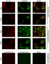
JAK2 kinase activity is required for ligand-induced EpoR colocalization with early endosomal marker EEA1. Cell-surface HA-EpoRs were labeled with anti-HA antibodies before Epo stimulation of 25 minutes. Cells were fixed and immunostained with anti-EEA1 antibodies followed by appropriate fluorescence-conjugated secondary antibodies. Representative confocal images (single section) for different conditions are presented. Images from negative controls with secondary antibodies alone are shown (original magnification ×40; Leica TCS SP5).
Figure 3
Figure 3
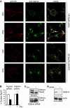
EpoR internalization is through a clathrin-dependent pathway. (A) γ2A cells expressing HA-EpoR with JAK2 or JAK2KD were seeded on glass coverslips. Cell-surface HA-EpoRs were labeled with anti-HA antibodies before Epo induction. Seven minutes after induction, cells were fixed and immunostained with anticlathrin antibodies followed by appropriate fluorescence-conjugated secondary antibodies. Representative confocal images (single section) are presented. Selected areas of colocalization are indicated with arrows (original magnification ×40; Leica TCS SP5). (B) Knockdown of the clathrin heavy chain abolished ligand-induced EpoR internalization. γ2A cells were transfected with siRNAs to the clathrin heavy chain, and surface EpoR was analyzed by flow cytometry. (C) Knockdown of the clathrin heavy chain abolished Epo-induced EpoR degradation. (D) Knockdown efficiency of the clathrin heavy chain shown by immunoblotting. Immunoblotting with actin was shown as a control.
Figure 4
Figure 4
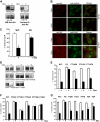
Y429, Y431, and Y479 mediate ligand-induced EpoR internalization. (A) Wild-type EpoR, but not F8, is degraded (left) on Epo stimulation. Wild-type EpoR, but not F8, is internalized on Epo stimulation as detected by surface IP (right). (B) F8 does not colocalize with clathrin or EEA1 on stimulation (original magnification ×40; Leica TCS SP5). (C) Epo-induced internalization of HA-F8 is dramatically reduced in TER119 erythroid progenitor cells from murine fetal livers. Surface expression of HA-EpoR or HA-F8 was measured 60 minutes after Epo stimulation. (D) Y429, Y431, or Y479 mediates ligand-induced EpoR internalization. Surface IP was performed on γ2A cells stably expressing HA-EpoR constructs with individual cytosolic tyrosine. (E,F) Y429, Y431, or Y479, but not other tyrosines, is sufficient for ligand-induced EpoR internalization. Ligand-induced receptor internalization was measured by flow cytometry in γ2A cells. (G) Replacing Y429, Y431, or Y479 individually on the wild-type EpoR (F429, F431, and F479) or replacing both Y429 and Y431 (Y2F) did not affect receptor internalization by flow cytometry, but simultaneously mutating all 3 tyrosines (Y3F) significantly reduced Epo-induced receptor internalization. *P = .025 (unpaired t test) versus control.
Figure 5
Figure 5
Concurrent knockdown of p85α and p85β diminishes receptor internalization. Receptor internalization was detected by flow cytometry in γ2A cells stably expressing EpoR and JAK2 and transfected with 100 nM siRNAs to SOCS3, p85α, p85β, or p85α and p85β together. Scrambled siRNA to GFP was used as a negative control. Immunoblots of each targeted protein demonstrate knockdown efficiency. Representative immunoblotting with actin was shown as a control. *P = .001 (unpaired t test) versus control.
Figure 6
Figure 6
p85α and p85β are important in mediating EpoR internalization. (A) Wortmannin treatment does not affect EpoR internalization. γ2A cells stably expressing HA-EpoR and JAK2 were treated with wortmannin for 2 hours at indicated concentrations followed by Epo stimulation. At 45 minutes after Epo stimulation, surface EpoRs were quantified by flow cytometry. Wortmannin inhibited AKT activation as detected by phospho-AKT antibodies. (B) Dominant-negative forms of p85α and p85β impair EpoR internalization. Epo-induced EpoR internalization was measured by flow cytometry in γ2A cells stably expressing HA-EpoR and JAK2 transiently transfected with vectors expressing the N or C terminal-SH2 domains from p85α and p85β. (1) vector control; (2) p85α N-terminal SH2; (3) p85α C-terminal SH2; (4) p85β N-terminal SH2; (5) p85β C-terminal SH2; (6) p85α and p85β N-terminal SH2s; (7) p85α and p85β C-terminal SH2s. *P = .001 (unpaired t test) versus control. (C) p85β binds EpoR on ligand stimulation. γ2A cells stably expressing HA-EpoR and JAK2 were transiently transfected with vectors expressing full-length p85β. At 48 hours after transfection, cells were starved overnight and treated with Epo for 10 minutes. Cell lysates were subjected to immunoprecipitation by p85β antibodies and immunoblotted with anti-HA antibody for the receptor. Cell lysates were also subjected to immunoblotting with antibodies to active JAK2 (P-JAK2), p85β, and HA. IP indicates immunoprecipitation; and IB, immunoblot.
Figure 7
Figure 7
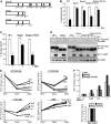
Truncated EpoR mutants fail to bind p85β or internalize on Epo stimulation. (A) Schematic diagram for truncated EpoR mutants used in our studies based on those from PFCP patients. (B) Truncated EpoR mutants did not internalize on Epo treatment by flow cytometry in γ2A cells. (C) Epo-induced internalization was defective for Stop2, but not Stop2-KYLYL, in erythroid progenitor cells. (D) Stop2+KYLYL, but not Stop1, Stop2, or Stop2+KFLFL, interacted with p85β on Epo induction. (E) Truncated EpoR mutants are hypersensitive to Epo in Ba/F3 cells. Ba/F3 cells stably expressing wild-type or truncated EpoR mutants were grown under different Epo concentrations. Cell numbers were measured by MTT assays every 24 hours for 3 days. Cell numbers from cultures in WEHI media as a source of IL-3 were shown as controls. (F) Erythroid progenitor cells expressing Stop2, but not Stop2+KYLYL, are hypersensitive to Epo. Erythroid progenitor cells were transduced with retroviruses expressing wild-type or truncated EpoR mutants. At 24 hours after infection, cells were washed and cultured in media containing different Epo concentration and 2% FBS (top panel), and cell numbers were measured by MTT assays after 24 hours. Cell numbers from cultures in media containing 10% FBS and 2 U/mL Epo (Epo media) were shown as controls (bottom panel). IP indicates immunoprecipitation; IB, immunoblot; P-JAK2, phosphorylated active JAK2.

References

    1. Constantinescu SN, Ghaffari S, Lodish HF. The erythropoietin receptor: structure, activation and intracellular signal transduction. Trends Endocrinol Metab. 1999;10:18–23. - PubMed
    1. Richmond TD, Chohan M, Barber DL. Turning cells red: signal transduction mediated by erythropoietin. Trends Cell Biol. 2005;15:146–155. - PubMed
    1. Klingmuller U, Wu H, Hsiao JG, et al. Identification of a novel pathway important for proliferation and differentiation of primary erythroid progenitors. Proc Natl Acad Sci U S A. 1997;94:3016–3021. - PMC - PubMed
    1. Bouscary D, Pene F, Claessens YE, et al. Critical role for PI 3-kinase in the control of erythropoietin-induced erythroid progenitor proliferation. Blood. 2003;101:3436–3443. - PubMed
    1. Zhao W, Kitidis C, Fleming MD, Lodish HF, Ghaffari S. Erythropoietin stimulates phosphorylation and activation of GATA-1 via the PI3-kinase/AKT signaling pathway. Blood. 2006;107:907–915. - PMC - PubMed

Publication types