. 2009 Apr 21;100(8):1320-9.
doi: 10.1038/sj.bjc.6605010. Epub 2009 Mar 31.

BCL6 degradation caused by the interaction with the C-terminus of pro-HB-EGF induces cyclin D2 expression in gastric cancers

Affiliations

BCL6 degradation caused by the interaction with the C-terminus of pro-HB-EGF induces cyclin D2 expression in gastric cancers

Y Hirata et al. Br J Cancer. .

Abstract

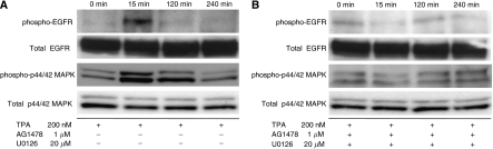
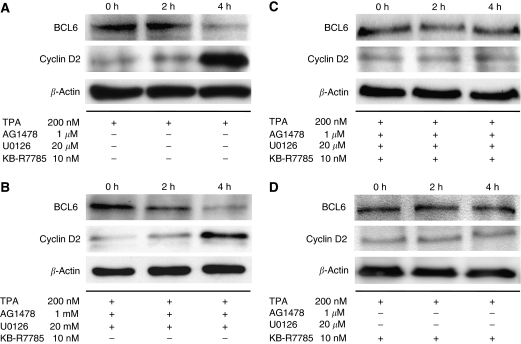
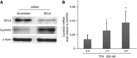
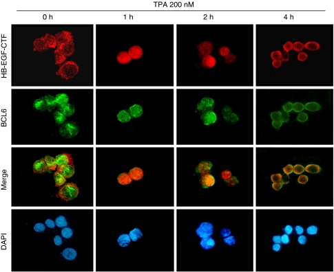
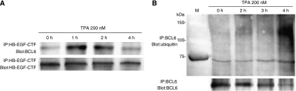
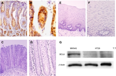
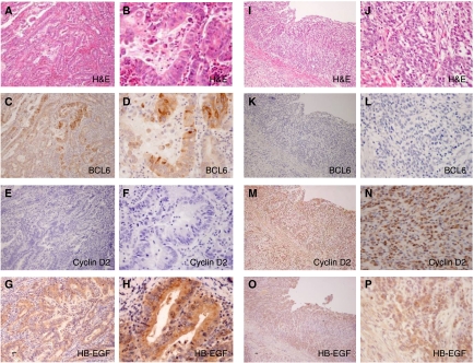
BCL6 is a transcriptional repressor that has important functions in lymphocyte differentiation and lymphomagenesis, but there have been no reports of BCL6 expression in gastric cancers. In the present study, we investigated the BCL6 function in gastric cancers. Treatment with TPA resulted in BCL6 degradation and cyclin D2 upregulation. This phenomenon was inhibited by the suppression of the nuclear translocation of HB-EGF-CTF (C-terminal fragment of pro-HB-EGF). The HB-EGF-CTF nuclear translocation leads to the interaction of BCL6 with HB-EGF-CTF and the nuclear export of BCL6, and after that BCL6 degradation was mediated by ubiquitin/proteasome pathway. Real-time RT-PCR and siRNA targeting BCL6 revealed that BCL6 suppresses cyclin D2 expression. Our data indicate that BCL6 interacts with nuclear-translocated HB-EGF-CTF and that the nuclear export and degradation of BCL6 induces cyclin D2 upregulation. We performed immunohistochemical analyses of BCL6, HB-EGF and cyclin D2 in human gastric cancers. The inverse correlation between BCL6 and cyclin D2 was also found in HB-EGF-positive human gastric cancers. BCL6 degradation caused by the HB-EGF-CTF also might induce cyclin D2 expression in human gastric cancers. Inhibition of HB-EGF-CTF nuclear translocation and maintenance of BCL6 function are important for the regulation of gastric cancer progression.


Figures

Figure 1
Figure 1
12-0-Tetradecanoylphorbor-13-acetate (TPA)-dependent ectodomain shedding induces phosphorylation of both EGFR and MAPK in MKN45 cells. (A) MKN45 cells were treated with 200 nM TPA for 0, 15, 120, and 240 min respectively. The endogenous phospho-EGFR and phospho-p44/42 MAPK, along with total EGFR and total p44/42 MAPK expression levels, were analysed by western blotting. (B) Before the treatment of MKN45 cells with 200 nM TPA for indicated periods, the cells were preincubated with 1 μM AG1478 (an EGFR tyrosine kinase inhibitor) and 20 μM U0126 (an MEK 1/2 inhibitor) for 60 min. The endogenous phospho-EGFR and phospho-p44/42 MAPK, along with total EGFR and total p44/42 MAPK expression levels, were analysed by western blotting.
Figure 2
Figure 2
Nuclear translocation of HB-EGF-CTF induces degradation of BCL6 and upregulation of cyclin D2 expression. (A) MKN45 cells were treated with 200 nM TPA for 2 or 4 h. The levels of BCL6 and cyclin D2 protein were determined by western blotting with anti-BCL6 and anti-cyclin D2 antibody. (B) Before the treatment of MKN45 cells with 200 nM TPA for 2 or 4 h, the cells were preincubated with 1 μM AG1478 and 20 μM U0126 for 60 min. The levels of BCL6 and cyclin D2 protein were analysed by western blotting. (C) Before TPA stimuli, 10 μM KB-R7785 (a metalloproteinase inhibitor) with both 1 μM AG1478 and 20 μM U0126 was added to the cells for 60 min. Then, MKN45 cells were treated with 200 nM TPA for 2 or 4 h. The levels of BCL6 and cyclin D2 protein were determined by western blotting. (D) Before TPA stimuli, 10 μM KB-R7785 alone was added to the cells for 60 min. Then, MKN45 cells were treated with 200 nM TPA for 2 or 4 h. The levels of BCL6 and cyclin D2 protein were determined by western blotting. The total incubation time from the start of the TPA treatment to cell lysis is shown at the top of the panel. β-Actin was used as a loading control for western blotting.
Figure 3
Figure 3
BCL6 suppresses cyclin D2 expression. (A) The levels of endogenous BCL6 and cyclin D2 protein were assessed by western blotting with anti-BCL6 and anti-cyclin D2 antibody, 48 h after transfection with siRNAs. (B) MKN45 cells were treated with 200 nM TPA for 1 or 3 h. The level of mRNA was analysed by real-time RT–PCR. The data are shown as means of three independent experiments. Bars, s.e. *P<0.01 vs no TPA (0 h).
Figure 4
Figure 4
Nuclear export of BCL6 is detected following nuclear translocation of HB-EGF-CTF after TPA treatment. HB-EGF-CTF (red) and BCL6 (green) were visualised by immunofluorescent microscopy. Nuclei were stained blue by DAPI. MKN45 cells were treated with 200 nM TPA for 4 h. The total incubation time is shown at the top of the panel.
Figure 5
Figure 5
BCL6 interacts with HB-EGF-CTF and is degraded through the ubiquitin/proteasome pathway. (A) After treatment with 200 nM TPA for the indicated periods, HB-EGF-CTF was immunoprecipitated from an extract of MKN45 cells with anti-HB-EGF-CTF antibody and subsequently divided into two equal aliquots. One aliquot was subjected to western blotting with an anti-BCL6 antibody, and the second aliquot was subjected to western blotting with anti-HB-EGF-CTF antibody to control for loading. (B) After treatment with 200 nM TPA for the indicated periods, BCL6 was immunoprecipitated from an extract of MKN45 cells with anti-BCL6 antibody and subsequently divided into two equal aliquots. One aliquot was subjected to western blotting with an anti-ubiquitin antibody, and the second aliquot was subjected to western blotting with anti-BCL6 antibody to control for loading. M, size markers.
Figure 6
Figure 6
BCL6 expression in human normal gastric epithelium and other human tissues. (A and B) Immunohistochemical staining of human normal gastric epithelium. (C and D) Immunohistochemical staining of human normal colon epithelium (E and F). Immunohistochemical staining of human normal oesophageal epithelium. (Original magnification: A × 100; C and E × 200; B, D, and F × 400). (G) The level of endogenous BCL6 protein of MKN45 cells, along with HT29 cells (human colon cancer cell line) and T.T (human oesophageal cancer cell line), was analysed by western blotting for comparison.
Figure 7
Figure 7
Immunohistochemistry of human gastric cancers. (AH) Differentiated gastric cancer and (IP) undifferentiated gastric cancer. (A and B) H&E staining; (C and D) BCL6 detected in the nucleus and cytoplasm of cancer cells; (E and F) cyclin D2 was negative; (G and H) HB-EGF was detected in the cytoplasm of cancer cells; (I and J) H&E staining; (K and L) BCL6 was negative; (M and N) cyclin D2 nuclear staining is observed in cancer cells; and (O and P) HB-EGF was detected in the cytoplasm of cancer cells. (Original magnification: A, C, E, G, I, K, M, O × 100; B, D, F, H, J, L, N, P ×400).

Similar articles

Cited by

References

    1. Alizadeh AA, Eisen MB, Davis RE, Ma C, Lossos IS, Rosenwald A, Boldrick JC, Sabet H, Tran T, Yu X, Powell JI, Yang L, Marti GE, Moore T, Hudson Jr J, Lu L, Lewis DB, Tibshirani R, Sherlock G, Chan WC, Greiner TC, Weisenburger DD, Armitage JO, Warnke R, Levy R, Wilson W, Grever MR, Byrd JC, Botstein D, Brown PO, Staudt LM (2000) Distinct types of diffuse large B-cell lymphoma identified by gene expression profiling. Nature 403: 503–511 - PubMed
    1. Asakura M, Kitakaze M, Takashima S, Liao Y, Ishikura F, Yoshinaka T, Ohmoto H, Node K, Yoshino K, Ishiguro H, Asanuma H, Sanada S, Matsumura Y, Takeda H, Beppu S, Tada M, Hori M, Higashiyama S (2002) Cardiac hypertrophy is inhibited by antagonism of ADAM12 processing of HB-EGF: metalloproteinase inhibitors as a new therapy. Nat Med 8: 35–40 - PubMed
    1. Barnard JA, Beauchamp RD, Russell WE, Dubois RN, Coffey RJ (1995) Epidermal growth factor-related peptides and their relevance to gastrointestinal pathophysiology. Gastroenterology 108: 564–580 - PubMed
    1. Baron BW, Nucifora G, McCabe N, Espinosa III R, Le Beau MM, McKeithan TW (1993) Identification of the gene associated with the recurring chromosomal translocations t(3;14)(q27;q32) and t(3;22)(q27;q11) in B-cell lymphomas. Proc Natl Acad Sci USA 90: 5262–5266 - PMC - PubMed
    1. Bos R, van Diest PJ, van der Groep P, Greijer AE, Hermsen MA, Heijnen I, Meijer GA, Baak JP, Pinedo HM, van der Wall E, Shvarts A (2003) Protein expression of B-cell lymphoma gene 6 (BCL-6) in invasive breast cancer is associated with cyclin D1 and hypoxia-inducible factor-1alpha (HIF-1alpha). Oncogene 22: 8948–8951 - PubMed

MeSH terms