Review
. 2009 May;10(5):285-90.
doi: 10.1038/nrg2571.

Genetic susceptibility to SLE: new insights from fine mapping and genome-wide association studies

Affiliations
Review

Genetic susceptibility to SLE: new insights from fine mapping and genome-wide association studies

Isaac T W Harley et al. Nat Rev Genet. 2009 May.

Abstract

Genome-wide association studies and fine mapping of candidate regions have rapidly advanced our understanding of the genetic basis of systemic lupus erythematosus (SLE). More than 20 robust associations have now been identified and confirmed, providing insights at the molecular level that refine our understanding of the involvement of host immune response processes. In addition, genes with unknown roles in SLE pathophysiology have been identified. These findings may provide new routes towards improved clinical management of this complex disease.


Figures

Figure 1
Figure 1. Pathways that contain established candidate SLE susceptibility loci
Genes involved in each pathway are indicated.
Figure 2
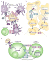
Figure 2. Pathways in which identified SLE risk alleles operate
Note that these are proposed models and it is possible, even likely, that the mechanism(s) by which these genes confer risk for SLE involve not only the pathways presented, but also other pathways. a | Phagocytosis. A presumed environmental trigger (for example, UV irradidation, viral infection or dysregulated apoptosis) leads to activation of antigen presenting cells which phagocytose self-antigen coated by opsonins (e.g. C3b), which are bound by their respective receptors (e.g. ITGAM). This leads to subsequent APC activation and presentation of self-antigen to host lymphocytes. This pathway potentially plays a role in disease initiation and perpetuation. In terms of initiation, a tendency towards antigen-presenting cell hyperactivation is a path toward loss of self-tolerance. In terms of perpetuation, when immune complexes are not cleared, this leads to the production of autoantibodies. [C1q: Complement component 1, subcomponent q; C2: Complement component 2; C3: Complement component 3; C3a: C3 cleavage product a; C3b: C3 cleavage product b; C4: Complement component 4; CRP: C-reactive protein; ITGAM: Intergrin αM; FcγR: Fc fragment of IgG receptor; HLA-DR: major histocompatibility complex, class II, DR] b | Type I interferon production: Recent data (48) suggest that TREX1 digests cytosolic DNA and prevents activation of a cell-intrinsic type I interferon (notably, interferon-α) response pathway (see 48). Similarly, activation of Toll-like receptors (TLRs – notably TLR7, TLR8, and TLR9) on ligand recognition (CpG DNA or ssRNA) leads to production of type I interferon by immune cells, notably plasmacytoid dendritic cells and the interferon responsive gene expression signature observed in lupus serum. [CpG DNA: DNA containing CpG dinucleotides; ssRNA: single-stranded RNA; TLR: toll-like receptor; IRAK1: Interleukin-1 receptor associated kinase-1; TNFAIP: Tumor necrosis factor-α induced protein 3 (aka A20); IRF5: interferon regulatory factor 5; IRF7: interferon regulatory factor 7; TREX1: three prime repair exonuclease 1; STAT4: signal transducer and activator of transcription 4; STAT1: signal transducer and activator of transcription 1] c | Immune Signal Transduction: Various stages in the life-cycle of lymphocytes are important for the development of the autoreactive B-cell clones, which produce the pathological autoantibodies observed in SLE. Here we focus on activation events. Self-antigen recognition by B-cells starts at the B-cell receptor (membrane IgM), where the balance of positive (B-cell receptor cross-linking) and negative signals (FCGR2B ligation) are transduced via intracellular kinases, such as BLK and BANK1, leading to B-cell activation. A similar process, leading to T cell activation occurs after uptake of the self-antigen and presentation on a class II MHC molecule, such as HLA-DR to a CD4+ T lymphocyte, which subsequently provides “Help” to B lymphocytes. It should be noted that autoreactive clones must avoid deletion before activation events can lead to florid autoimmunity. [BCR: B cell receptor; FCGR2B: Fc fragment of IgG receptor, low affinity 2B; BLK: B lymphoid tyrosine kinase; BANK1: B-cell scaffold protein with ankyrin repeats 1; TNFRSF4: tumor necrosis factor receptor superfamily, member 4; TNFSF4: tumor necrosis factor superfamily, member 4; PTPN22: protein tyrosine phosphatase, non-receptor type 22; TCR: T cell receptor; PDCD1: programmed cell death 1; CD274: programmed cell death 1 ligand 1 precursor; PDCD1LG2: programmed cell death 1 ligand 2 precursor].

References

    1. Lau CS, Yin G, Mok MY. Ethnic and geographical differences in systemic lupus erythematosus: an overview. Lupus. 2006;15:713–714. - PubMed
    1. Lockshin MD. Sex differences in autoimmune disease. Lupus. 2006;15:753–756. - PubMed
    1. Uribe AG, McGwin G, Jr, Reveille JD, Alarcon GS. What have we learned from a 10-year experience with the LUMINA (Lupus in Minorities; nature vs Nurture) cohort? Where are we heading? Autoimmun Rev. 2004;3:321–329. - PubMed
    1. Hochberg MC. Updating the American College of Rheumatology revised criteria for the classification of systemic lupus erythematosus. Arthritis Rheum. 1997;40:1725. - PubMed
    1. Tan EM, Cohen AS, Fries JF, Masi AT, McShane DJ, et al. The 1982 revised criteria for the classification of systemic lupus erythematosus. Arthritis Rheum. 1982;25:1271–1277. - PubMed

Publication types

MeSH terms