. 2009;4(4):e5060.
doi: 10.1371/journal.pone.0005060. Epub 2009 Apr 1.

The synovial sarcoma-associated SYT-SSX2 oncogene antagonizes the polycomb complex protein Bmi1

Affiliations

The synovial sarcoma-associated SYT-SSX2 oncogene antagonizes the polycomb complex protein Bmi1

Roy Barco et al. PLoS One. 2009.

Abstract

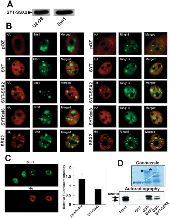
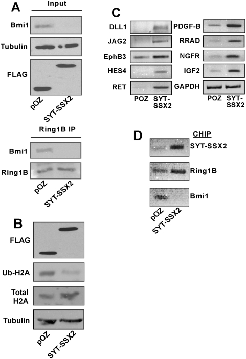
This study demonstrates deregulation of polycomb activity by the synovial sarcoma-associated SYT-SSX2 oncogene, also known as SS18-SSX2. Synovial sarcoma is a soft tissue cancer associated with a recurrent t(X:18) translocation event that generates one of two fusion proteins, SYT-SSX1 or SYT-SSX2. The role of the translocation products in this disease is poorly understood. We present evidence that the SYT-SSX2 fusion protein interacts with the polycomb repressive complex and modulates its gene silencing activity. SYT-SSX2 causes destabilization of the polycomb subunit Bmi1, resulting in impairment of polycomb-associated histone H2A ubiquitination and reactivation of polycomb target genes. Silencing by polycomb complexes plays a vital role in numerous physiological processes. In recent years, numerous reports have implicated gain of polycomb silencing function in several cancers. This study provides evidence that, in the appropriate context, expression of the SYT-SSX2 oncogene leads to loss of polycomb function. It challenges the notion that cancer is solely associated with an increase in polycomb function and suggests that any imbalance in polycomb activity could drive the cell toward oncogenesis. These findings provide a mechanism by which the SYT-SSX2 chimera may contribute to synovial sarcoma pathogenesis.


Conflict of interest statement

Competing Interests: The authors have declared that no competing interests exist.

Figures

Figure 1
Figure 1. SYT-SSX2 associates with Polycomb complex proteins.
(A) Equivalent protein levels of retrovirally expressed SYT-SSX2 (FLAG and HA-tagged) in U2-OS cells and in a primary human synovial sarcoma cell line (Syn1). 100 ug of cellular lysate-derived protein was loaded in each lane. SYT-SSX2 was visualized with the anti-FLAG tag antibody in U2-OS and with a SYT-specific antibody [SV11; 37] in Syn1 cells. (B) Colocalization of SYT-SSX2 with Bmi1 and Ring1B. Cells infected with pOZ viral vector, SYT, SYT-SSX2, SYTdel8, and SSX2 (all FLAG and HA-tagged), were analyzed by indirect immunofluorescence. Infected cDNAs (HA, red) and Bmi1 or Ring1B (green) were visualized individually and with merging of the two channels (merge). The cytoplasmic HA staining in pOZ-infected cells is due to the generation of an irrelevant FLAG/HA-tagged peptide by the vector. Image magnification was at 63×. (C) Decrease in Bmi1 fluorescence in SYT-SSX2 expressing cells. U2OS cells infected with SYT-SSX2 were analyzed by indirect immunofluorescence for SYT-SSX2 (HA; red) and Bmi (green). Image magnification was at 63×. Average fluorescence (20 cells per replicate; n = 3) of Bmi1 was compared between SYT-SSX2-infectants and uninfected cells using MetaMorph software and plotted. (D) In vitro binding of Ring1B to SYT-SSX2. Upper panel: GST, GST-Bmi1 and GST-SYT-SSX2 visualized by Coomassie staining. The asterisks (*) indicate full-length proteins. Lower panel: autoradiography of in vitro-translated (IVT) Ring1B bound to GST-Bmi1 (positive control) and GST-SYT-SSX2. Lane 1 represents 10% of input IVT Ring1B.
Figure 2
Figure 2. Loss of Bmi1 immunoreactivity following de novo expression of SYT-SSX2.
(A) Extracts derived from pOZ vector, SYT and SYT-SSX2 cells were analyzed by Western blotting for levels of Bmi1 and alpha-tubulin (loading control). (B) Loss of immunoreactivity is specific to Bmi1. pOZ and SYT-SSX2-infectants were lysed and immunoblotted for Bmi1, Ring1B, YY1, FLAG (to detect ectopically expressed proteins) and alpha-tubulin. (C) The decrease in Bmi1 signal is a specific function of SYT-SSX2 chimera. Extracts from uninfected cells, pOZ, SYT, SYT-SSX2, SYTdel8 and SSX2 were immunoblotted for Bmi1, FLAG-tagged ectopically expressed proteins and alpha-tubulin. The FLAG-tagged protein visualized in pOZ-infected cells is due to the generation of an irrelevant FLAG/HA-tagged peptide by the vector. The molecular weight of the SSX2 domain is ∼10 Kd. For its detection we ran the SSX2 lysates on a separate 18% SDS-PAGE system. The remaining lysates were resolved on a 10% SDS-PAGE. U: uninfected cells.
Figure 3
Figure 3. Mapping of the Polycomb association region on SYT-SSX2.
(A) Schematic of wildtype SYT-SSX2 and the truncation mutants generated within the SSX2 region. (B) Immunofluorescent colocalization studies of the SYT-SSX2 truncation mutants (HA, red) and Bmi1 (green), with merging of the two channels displayed as well. Also indicated is the average percentage of SYT-SSX2 (or mutant) proteins aggregating with polycomb bodies (a minimum of 100 polycomb foci counted per replicate; n = 3). Image magnification was at 63×. (C) Plot of Bmi1 fluorescence in U2OS cells expressing the SXdel mutants and the POZ control vector. Average fluorescence (20 cells per replicate; n = 3) of Bmi1 was compared between POZ- and SXdel-infectants. The Bmi1 fluorescence in the POZ nuclei was consistently equivalent to that of uninfected cells. Data analysis was performed using MetaMorph software. (D) Loss of Bmi1 immunoreactivity correlates with association of SYT-SSX2 to polycomb complexes. Western blotting of cells infected with indicated FLAG-tagged proteins using Bmi1, FLAG and alpha-tubulin-specific antibodies.
Figure 4
Figure 4. Loss of Bmi1 immunoreactivity in SYT-SSX2-infected cells results from depletion of the Bmi1 protein.
(A) Real time RT-PCR of Bmi1 in U2OS lysates expressing the indicated proteins. (B) Left panel: overexpressed polyoma epitope-tagged Bmi1 (2PY-Bmi1) was infected with either SYT-SSX2 or pOZ backbone. Cell lysates were immunoblotted with Bmi1 antibody, then stripped and reprobed with an anti polyoma tag antibody. Immunoblotting for alpha-tubulin was used as a loading control. Right panel: cell lysates of pOZ and SYT-SSX2-infected cells were immunoblotted for either mouse monoclonal or rabbit polyclonal Bmi1 antibody. Immunoblotting for alpha-tubulin was used as a loading control. (C) Densitometry plotting of pulse-chase analysis of 2PY-Bmi1 in pOZ and SYT-SSX2-infected cells. 2PY-Bmi1-expressing cells infected with pOZ and SYT-SSX2 were labeled for 1 hr. with S35–labeled Methionine and Cysteine. Labeled cells were chased at the indicated timepoints and immunoprecipitated with a polyoma tag antibody. Immunoprecipitated 2PY-Bmi1 band intensities were quantitated by densitometry and plotted with pOZ and SYT-SSX2 experiments indicated. Pulse-chase experiment was successfully replicated (n = 2). (D) Immunoprecipitation studies demonstrating the specificity of the immunoprecipitated 2PY-Bmi1 band used in pulse-chase analysis. Negative controls using polyoma peptide blocking (lane 3), no antibody (lane 4) and naïve U2OS cells (lane 1) are indicated.
Figure 5
Figure 5. Functional antagonism of Bmi1 in SYT-SSX2-expressing cells.
(A) Lower panel: the Ring1B-interacting Bmi1 population is depleted in SYT-SSX2-expressing cells. pOZ and SYT-SSX2 infected cell extracts were immunoprecipitated (IP) with a Ring1B antibody. Immunoprecipitates were immunoblotted with the antibodies indicated to the left. FLAG antibody was used to detect ectopically expressed SYT-SSX2 (upper panel). (B) Extracts from pOZ and SYT-SSX2 infectants were immunoblotted with anti-ubiquityl H2A, total H2A and FLAG antibodies. Immunoblotting for alpha-tubulin was used as a loading control. (C) Increased transcription of SYT-SSX2-derepressed polycomb targets. Relative RT-PCR of a subset of SYT-SSX2-upregulated genes also shown to be polycomb-silenced targets . GAPDH served as loading control. D) SYT-SSX2 (upper image), Ring 1B (middle image) and Bmi1 (lower image) occupancy on the NGFR promoter by CHIP-PCR in pOZ vector- and SYT-SSX2-expressing U2-OS cells.

References

    1. Sparmann A, van Lohuizen M. Polycomb silencers control cell fate, development and cancer. Nat Rev Cancer. 2006;6:846–856. - PubMed
    1. Ringrose L, Paro R. Epigenetic regulation of cellular memory by the Polycomb and Trithorax group proteins. Annu Rev Genet. 2004;38:413–443. - PubMed
    1. van der Vlag J, Otte AP. Transcriptional repression mediated by the human polycomb-group protein EED involves histone deacetylation. Nat Genet. 1999;23:474–478. - PubMed
    1. Viré E, Brenner C, Deplus R, Blanchon L, Fraga M, et al. The Polycomb group protein EZH2 directly controls DNA methylation. Nature. 2006;439:871–874. - PubMed
    1. Czermin B, Melfi R, McCabe D, Seitz V, Imhof A, Pirrotta V. Drosophila enhancer of Zeste/ESC complexes have a histone H3 methyltransferase activity that marks chromosomal Polycomb sites. Cell. 2002;111:185–196. - PubMed

Publication types

MeSH terms