. 2009;4(4):e5052.
doi: 10.1371/journal.pone.0005052. Epub 2009 Apr 1.

VEGFA upregulates FLJ10540 and modulates migration and invasion of lung cancer via PI3K/AKT pathway

Affiliations

VEGFA upregulates FLJ10540 and modulates migration and invasion of lung cancer via PI3K/AKT pathway

Chang-Han Chen et al. PLoS One. 2009.

Abstract

Background: Lung adenocarcinoma is the leading cause of cancer-related deaths among both men and women in the world. Despite recent advances in diagnosis and treatment, the mortality rates with an overall 5-year survival of only 15%. This high mortality is probably attributable to early metastasis. Although several well-known markers correlated with poor/metastasis prognosis in lung adenocarcinoma patients by immunohistochemistry was reported, the molecular mechanisms of lung adenocarcinoma development are still not clear. To explore novel molecular markers and their signaling pathways will be crucial for aiding in treatment of lung adenocarcinoma patients.

Methodology/principal findings: To identify novel lung adenocarcinoma-associated /metastasis genes and to clarify the underlying molecular mechanisms of these targets in lung cancer progression, we created a bioinformatics scheme consisting of integrating three gene expression profile datasets, including pairwise lung adenocarcinoma, secondary metastatic tumors vs. benign tumors, and a series of invasive cell lines. Among the novel targets identified, FLJ10540 was overexpressed in lung cancer tissues and is associated with cell migration and invasion. Furthermore, we employed two co-expression strategies to identify in which pathway FLJ10540 was involved. Lung adenocarcinoma array profiles and tissue microarray IHC staining data showed that FLJ10540 and VEGF-A, as well as FLJ10540 and phospho-AKT exhibit positive correlations, respectively. Stimulation of lung cancer cells with VEGF-A results in an increase in FLJ10540 protein expression and enhances complex formation with PI3K. Treatment with VEGFR2 and PI3K inhibitors affects cell migration and invasion by activating the PI3K/AKT pathway. Moreover, knockdown of FLJ10540 destabilizes formation of the P110-alpha/P85-alpha-(PI3K) complex, further supporting the participation of FLJ10540 in the VEGF-A/PI3K/AKT pathway.

Conclusions/significance: This finding set the stage for further testing of FLJ10540 as a new therapeutic target for treating lung cancer and may contribute to the development of new therapeutic strategies that are able to block the PI3K/AKT pathway in lung cancer cells.


Conflict of interest statement

Competing Interests: The authors have declared that no competing interests exist.

Figures

Figure 1
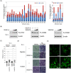
Figure 1. FLJ10540 exhibits differentially expressed patterns in lung adenocarcinomas, metastatic tumors, and a series of human lung invasive cancer cell lines. (CL1-0, CL1-1, CL1-5, and CL1-5-F4, in the order of increasing invasive activity).
(A, left panel) The microarray expression patterns of FLJ10540 in lung adenocarcinoma patients were normalized against the expression patterns on HG_U133A chips. N: adjacent non-tumor tissues; T: tumor tissues. The boxplot shows the data distribution as a grouping classification and indicates that there is a statistically significant difference (p<0.0001) between the tumor tissues and the adjacent nont-umor tissues from the same lung cancer patient. (A, right panel) The mRNA levels of FLJ10540 in samples from 16 lung cancer patients were determined by Q-RT-PCR. The results were normalized against the expression levels of GAPDH mRNA in each paired sample and plotted with boxplot. (B) The microarray expression patterns of FLJ10540 were compared among 30 benign tumors, 20 tumor metastases to the lymph node, and 200 metastatic tumors, and were showed with boxplot. (C, left panel) The mRNA levels of FLJ10540 were determined by Q-RT-PCR in lung cancer cell lines. The results were normalized against the level of GAPDH mRNA in each cell line. The experiments were performed in triplicate. (C, right panel) Total protein was extracted from CL1-0 and CL1-5-F4 cells and subjected to immunoblot analysis with anti-FLJ10540. β-actin was used an internal loading control.
Figure 2
Figure 2. FLJ10540 overexpression promotes lung cancer cell migration and invasion.
(A, left panel) HA-tagged-FLJ10540 stable clones of CL1-0 cells were established. The cell lysates were subjected to immunoblot analysis with anti-HA antibody. (A, middle panel) Phase-contrast images of monolayer cultures of CL1-0 cells expressing FLJ10540 and vehicle control were shown. (A, right panel) To measure cell growth rates, 5×10³ cells of vehicle-CL1-0 and CL1-0-FLJ10540 stable clones were plated at day 0 in 96-well plates with 10% FBS. The cells were counted at indicated time points by the MTT assay (OD570) to quantify the cell growth. Data were normalized against the OD570 value on day 0. The growth rate of CL1-0 cells is shown as the mean±s.d. of three independent experiments. (B, upper panel) For the migration assay, 5×10³ cells of vehicle-CL1-0 and CL1-0-FLJ10540 stable clones were seeded into the top of a Transwell insert. After 24 hours, the cells on the topside were scraped, and the cells that migrated to the bottom were fixed and stained with Giemsa. The migration relative-fold of vehicle-CL1-0 and CL1-0-FLJ10540 stable clones was normalized with vehicle control and presented diagrammatically. (B, bottom panel) For the invasion assay, 1×10⁴ cells were seeded after Matrigel was added. The invasion relative-fold of stable clone was normalized against vehicle cells and represented diagrammatically. All of the data represent the mean±s.d. of three independent experiments.
Figure 3
Figure 3. The migration and invasion abilities of lung cancer cells are inhibited by FLJ10540-mediated siRNAs.
(A and B, left panel) A negative control siRNA with two different FLJ10540 siRNAs (siFLJ10540-1 and siFLJ10540-2) were transfected into CL1-5 and H1299 cells for 24 hours. After transfection, western blotting was performed using anti-FLJ10540 and anti-β-actin antibodies. (A and B, middle panel) To measure cell growth rates, 5×10³ cells of negative control-CL1-5, CL1-5-FLJ10540 siRNAs, negative control-H1299, and H1299-FLJ10540 siRNAs were plated at day 0 in 96 well plates with 10% FBS. The cells were counted at indicated time points by the MTT assay (OD570) to quantify the cell growth. Data were normalized against the OD570 value on day 0 of each treatment. The growth rate of CL1-5 and H1299 cells are shown as the mean±s.d. of three independent experiments. (A and B, right panel) The migration relative-fold and the invasion relative-fold of CL1-5 and H1299 were normalized against negative control cells and represented diagrammatically. The results represent the mean±s.d. of three independent experiments.
Figure 4
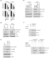
Figure 4. VEGF-A promotes FLJ10540 protein expression and enhances FLJ10540-induced migration and invasion in lung cancer cells.
(A) The microarray expression patterns of VEGF-A and FLJ10540 in lung adenocarcinoma patients were shown. The results were normalized against the expression patterns of 56 chips (HG_U133A). N: adjacent non-tumor tissues; T: tumor tissues. (B, left panel) VEGF-A induced an increase in FLJ10540 protein levels in a dose-dependent manner. After treatment with various concentration of VEGF-A (left panel) or VEGF-B (right panel) for 10 min in CL1-0 cells, the total proteins were extracted from CL1-0 cells and probed with antibody against FLJ10540. (B, middle panel) Serum-starved CL1-0 cells were pre-treated with or without various concentrations of SU5416 for 2 hours; cells were then stimulated with 20 ng/ml VEGF-A for 10 min. β-actin was used as an internal loading control. (C) Serum-starved CL1-0 cells were pre-treated with or without SU5416 for 2 hours; cells were then stimulated with VEGF-A (at the concentration of 20 ng/ml) for 3 hours. The migration and invasion relative-folds were normalized against vehicle cells. (D, left, upper panel) For the migration assay, 5×10³ cells of vehicle-CL1-0 and CL1-0-FLJ10540 stable clones were seeded into the top of a Transwell insert and allowed to adhere for 12 hours, and were then incubated with or without VEGF-A (20 ng/ml) for 3 hours. At the end of the assay, the cells on the topside were scraped, and the cells that migrated to the bottom were fixed and stained with Giemsa. (D, left, bottom panel) For the invasion assay, 1×10⁴ cells were seeded after Matrigel was added, and were then incubated with or without VEGF-A (20 ng/ml) for 3 hours. All of the data represent the mean±s.d. of three independent experiments. (E) Indirect immunofluorescence analysis of FLJ10540 in VEGF-A-treated cells. The protein expression and the subcellular localization of FLJ10540 were analyzed in the presence or absence of VEGF-A (20 ng/ml) in CL1-0 cells using immunofluorescence microscopy. After being incubated with or without VEGF-A for 30 min or 180 min, the cells were fixed with 3.7% formaldehyde and processed for indirect immunofluorescence microscopy. FLJ10540 translocated to the cell membrane upon VEGF-A treatment (Arrowhead). Bar: 10 µm.
Figure 5
Figure 5. Histopathologic and immunophenotypic findings in lung adenocarcinomas with immunoreactivity for VEGF-A, FLJ10540, and phosphorylated AKT.
(A) The tumor has a papillary growth pattern (Hematoxylin & Eosin, 100×). (B) The tumor cells are immunoreactive with VEGF-A, with a staining intensity score of 2 (100×). (C) The tumor cells are immunoreactive with FLJ10540, with a staining intensity score of 2 (100×). (D) The tumor cells are immunoreactive with phosphorylated AKT, with a staining intensity score of 3 (100×).
Figure 6
Figure 6. FLJ10540 alone or increased FLJ10540, mediated by VEGF-A stimulation, not only modulates cell migration and invasion through the PI3K/AKT signaling pathway, but also reinforces PI3K complex formation.
(A) Vehicle-CL1-0, CL1-0-FLJ10540, vehicle-RK3E, and RK3E-FLJ10540 infected cells were serum-starved for 24 hours and treated with or without the indicated inhibitors, including SB202190, PD98059, and LY294002 for 2 hours. The migration and invasion ratios of vehicle-CL1-0, CL1-0-HA-FLJ10540, vehicle-RK3E, and RK3E-Flag-FLJ10540 infected cells were determined as previously described. (B) CL1-0 and RK3E-expressing FLJ10540 cells were treated with or without LY294002 at the final concentration of 10 µM. The total cell lysates were subjected to immunoblot analysis for the unphosphorylated or phosphorylated form of AKT. β-actin was used as an internal loading control. (C) CL1-0 cells expressing HA-FLJ10540 were pre-treated with or without VEGF-A (20 ng/ml). After 10 min, LY294002 (10 µM) was added and cells were further incubated for 2 hours. The total cell lysates were subjected to immunoblot analysis for HA, FLJ10540, or the unphosphorylated or phosphorylated forms of AKT. (D) A negative control siRNA and two different FLJ10540 siRNAs (siFLJ10540-1 and siFLJ10540-2) were transfected into CL1-0 cells for 24 hours. After transfection, cells were treated with or without VEGF-A (20 ng/ml) for 10 min. Western blotting was performed as in (C). (E upper panel) Serum-starved CL1-0 cells were pre-treated with or without SU5416 for 2 hours; cells were then stimulated with or without 20 ng/ml VEGF-A for 10 min. Cell lysates were immunoprecipitated with polyclonal antibodies against p110-α or protein IgG (as a control), which was followed by immunoblotting with p110-α, p85-α, and FLJ10540. (E lower panel) A negative control siRNA and two different FLJ10540 siRNAs (siFLJ10540-1 and siFLJ10540-2) were transfected into CL1-0 cells for 24 hours. After transfection, cell lysates were immunoprecipitated with polyclonal antibodies against p110-α, p85-α, or protein IgG (as a control), followed by immunoblotting with p110-α, p85-α, and FLJ10540.

Similar articles

Cited by

References

    1. Parkin DM, Bray F, Ferlay J, Pisani P. Global cancer statistics, 2002. CA Cancer J Clin. 2005;55:74–108. - PubMed
    1. Jemal A, Siegel R, Ward E, Hao Y, Xu J, et al. Cancer statistics, 2008. CA Cancer J Clin. 2008;58:71–96. - PubMed
    1. GA KBaP. Surgical management of non-small cell lung cancer. Pearson's Thoracic & Esophageal Surgery. Philadelphia, PA: Churchill Livingstone/ Elsevier; 2008. pp. 765–780.
    1. Friedl P, Wolf K. Tumour-cell invasion and migration: diversity and escape mechanisms. Nat Rev Cancer. 2003;3:362–374. - PubMed
    1. Hendrix MJ, Seftor EA, Chu YW, Trevor KT, Seftor RE. Role of intermediate filaments in migration, invasion and metastasis. Cancer Metastasis Rev. 1996;15:507–525. - PubMed

Publication types

MeSH terms