

New imaging techniques in the diagnosis of multiple sclerosis
- PMID: 19337386
- PMCID: PMC2662586
- DOI: 10.1517/17530050802361161
New imaging techniques in the diagnosis of multiple sclerosis
Abstract
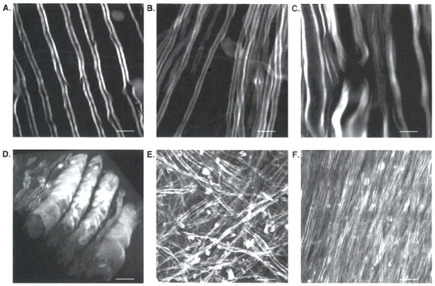
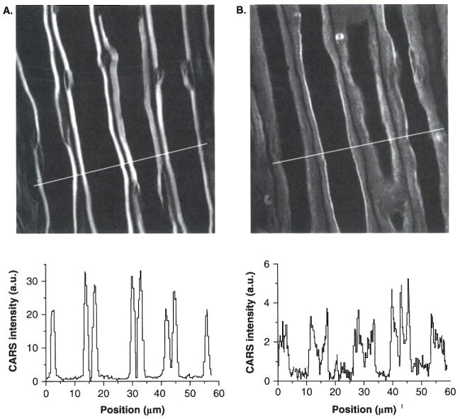
Background: Multiple sclerosis (MS) is a chronic disabling disorder histopathologically characterized by inflammation, demyelination and axonal loss. Conventional MRI has made most contributions to the diagnosis of MS. However, it is not sufficiently sensitive and specific to reveal the extent and severity of the damage in the disease. Other nuclear magnetic resonance (NMR) techniques including magnetic resonance spectroscopy, magnetization transfer imaging, diffusion weighted and diffusion tensor imaging, and functional MRI have provided additional information that improves the diagnosis and understanding of MS. Optical techniques including optical coherence tomography (OCT) and coherent anti-Stokes Raman scattering (CARS) microscopy have shown promise in diagnosis and mechanistic study of myelin diseases.
Objective: To review new imaging techniques and their potential in diagnosis of MS.
Method: The principles of three imaging techniques (MRI, OCT and CARS) and their applications to MS studies are described. Their advantages and disadvantages are compared.
Conclusion: Conventional MRI remains a critical tool in the diagnosis of MS. Alternative NMR/MRI techniques have improved specificity for the detection of lesions and provided more quantitative information about MS. Optical techniques including OCT and CARS microscopy are opening up new ways for diagnosis and mechanistic study of myelin diseases.
Keywords: coherent anti-Stokes Raman scattering microscopy; magnetic resonance imaging; multiple sclerosis; optical coherence tomography.
Figures
References
-
- Compston A, Coles A. Multiple sclerosis. Lancet. 2002;359:1221–31. - PubMed
-
- Bagnato F, Jeffries N, Richert ND, et al. Evolution of T1 black holes in patients with multiple sclerosis imaged monthly for 4 years. Brain. 2003;126:1782–9. - PubMed
-
- Brück W, Bitsch A, Kolenda H, et al. Inflammatory central nervous system demyelination: correlation of magnetic resonance imaging findings with lesion pathology. Ann Neurol. 1997;42:783–93. - PubMed
-
- McDonald WI, Compston A, Edan G, et al. Recommended diagnostic criteria for multiple sclerosis: guidelines from the international panel on the diagnosis of multiple sclerosis. Ann Neurol. 2001;50:121–7. - PubMed
-
- Filippi M, Grossman RI. MRI techniques to monitor MS evolution: the present and the future. Neurology. 2002;58:1147–53. - PubMed
Grants and funding
LinkOut - more resources
Full Text Sources