Bipolar disorder and neurophysiologic mechanisms
- PMID: 19337455
- PMCID: PMC2646644
- DOI: 10.2147/ndt.s4329
Bipolar disorder and neurophysiologic mechanisms
Abstract
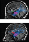
Recent studies have suggested that some variants of bipolar disorder (BD) may be due to hyperconnectivity between orbitofrontal (OFC) and temporal pole (TP) structures in the dominant hemisphere. Some initial MRI studies noticed that there were corpus callosum abnormalities within specific regional areas and it was hypothesized that developmentally this could result in functional or effective connectivity changes within the orbitofrontal-basal ganglia-thalamocortical circuits. Recent diffusion tensor imaging (DTI) white matter fiber tractography studies may well be superior to region of interest (ROI) DTI in understanding BD. A "ventral semantic stream" has been discovered connecting the TP and OFC through the uncinate and inferior longitudinal fasciculi and the elusive TP is known to be involved in theory of mind and complex narrative understanding tasks. The OFC is involved in abstract valuation in goal and sub-goal structures and the TP may be critical in binding semantic memory with person-emotion linkages associated with narrative. BD patients have relative attenuation of performance on visuoconstructional praxis consistent with an atypical localization of cognitive functions. Multiple lines of evidence suggest that some BD alleles are being selected for which could explain the enhanced creativity in higher-ability probands. Associations between ROI's that are not normally connected could explain the higher incidence of artistic aptitude, writing ability, and scientific achievements among some mood disorder subjects.
Keywords: artistic aptitude; bipolar disorder; creativity; diffusion tensor imaging; inferior fronto-occipital fasciculus; inferior longitudinal fasciculus; mood dysphoria; uncinate fasciculus; ventral semantic stream; white matter tractography; writing ability.
Figures


Similar articles
-
Language networks in semantic dementia.Brain. 2010 Jan;133(Pt 1):286-99. doi: 10.1093/brain/awp233. Epub 2009 Sep 16. Brain. 2010. PMID: 19759202 Free PMC article.
-
Independent contribution of individual white matter pathways to language function in pediatric epilepsy patients.Neuroimage Clin. 2014 Sep 30;6:327-32. doi: 10.1016/j.nicl.2014.09.017. eCollection 2014. Neuroimage Clin. 2014. PMID: 25379446 Free PMC article.
-
Identifying preoperative language tracts and predicting postoperative functional recovery using HARDI q-ball fiber tractography in patients with gliomas.J Neurosurg. 2016 Jul;125(1):33-45. doi: 10.3171/2015.6.JNS142203. Epub 2015 Dec 11. J Neurosurg. 2016. PMID: 26654181
-
White matter modifications of corpus callosum in bipolar disorder: A DTI tractography review.J Affect Disord. 2023 Oct 1;338:220-227. doi: 10.1016/j.jad.2023.06.012. Epub 2023 Jun 9. J Affect Disord. 2023. PMID: 37301293 Review.
-
Cognitive deficits in bipolar disorders: Implications for emotion.Clin Psychol Rev. 2018 Feb;59:126-136. doi: 10.1016/j.cpr.2017.11.006. Epub 2017 Nov 21. Clin Psychol Rev. 2018. PMID: 29195773 Free PMC article. Review.
Cited by
-
Multisensory integration processing during olfactory-visual stimulation-An fMRI graph theoretical network analysis.Hum Brain Mapp. 2018 Sep;39(9):3713-3727. doi: 10.1002/hbm.24206. Epub 2018 May 7. Hum Brain Mapp. 2018. PMID: 29736907 Free PMC article.
-
Identification and individualized prediction of clinical phenotypes in bipolar disorders using neurocognitive data, neuroimaging scans and machine learning.Neuroimage. 2017 Jan 15;145(Pt B):254-264. doi: 10.1016/j.neuroimage.2016.02.016. Epub 2016 Feb 13. Neuroimage. 2017. PMID: 26883067 Free PMC article.
-
Intuition, insight, and the right hemisphere: Emergence of higher sociocognitive functions.Psychol Res Behav Manag. 2010;3:1-39. doi: 10.2147/prbm.s7935. Epub 2010 Mar 3. Psychol Res Behav Manag. 2010. PMID: 22110327 Free PMC article.
-
DTI tractography and white matter fiber tract characteristics in euthymic bipolar I patients and healthy control subjects.Brain Imaging Behav. 2013 Jun;7(2):129-39. doi: 10.1007/s11682-012-9202-3. Brain Imaging Behav. 2013. PMID: 23070746 Free PMC article.
-
Initial and Progressive Gray Matter Abnormalities in Insular Gyrus and Temporal Pole in First-Episode Schizophrenia Contrasted With First-Episode Affective Psychosis.Schizophr Bull. 2016 May;42(3):790-801. doi: 10.1093/schbul/sbv177. Epub 2015 Dec 16. Schizophr Bull. 2016. PMID: 26675295 Free PMC article.
References
-
- Aboitiz F, Scheibel AB, Fisher RS, et al. Fiber composition of the human corpus callosum. Brain Res. 1992;598:143–53. - PubMed
-
- Alexander GE, DeLong MR, Strick PL. Parallel organization of functionally segregated circuits linking basal ganglia and cortex. Annu Rev Neurosci. 1986;9:357–81. - PubMed
-
- Ali SO, Denicoff KD, Altshuler LL, et al. A preliminary study of the relation of neuropsychological performance to neuroanatomic structures in bipolar disorder. Neuropsy Neuropsy Be. 2000;13:20–8. - PubMed
-
- Anderson AK, Christoff K, Stappen I, et al. Dissociated neural representations of intensity and valence in human olfaction. Nat Neurosci. 2003;6:196–202. - PubMed
Grants and funding
LinkOut - more resources
Full Text Sources

