. 2009 Oct;27(10):1298-305.
doi: 10.1002/jor.20886.

Prolyl hydroxylase inhibitors increase neoangiogenesis and callus formation following femur fracture in mice

Affiliations

Prolyl hydroxylase inhibitors increase neoangiogenesis and callus formation following femur fracture in mice

Xing Shen et al. J Orthop Res. 2009 Oct.

Abstract

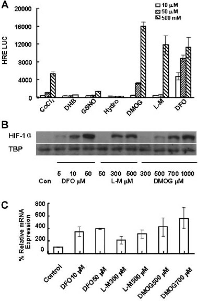
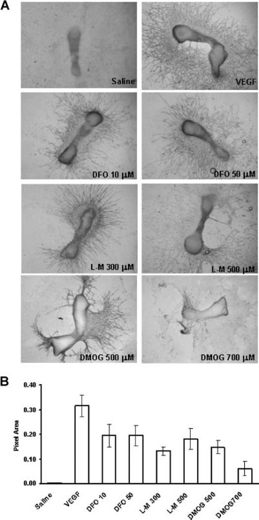
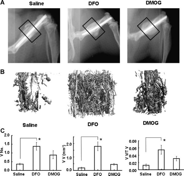
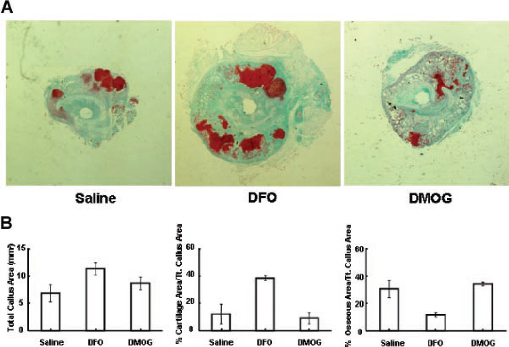
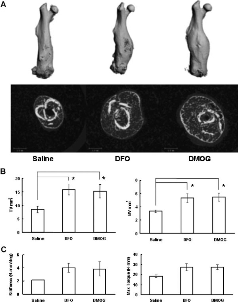
Skeletal trauma and impaired skeletal healing is commonly associated with diminished vascularity. Hypoxia inducible factor alpha (HIF-1) is a key transcription factor responsible for activating angiogenic factors during development and tissue repair. Small molecule inhibitors of the prolyl hydroxylase enzyme (PHD), the key enzyme responsible for degrading HIF-1, have been shown to activate HIF-1, and are effective in inducing angiogenesis. Here we examined the effects of several commercially available PHD inhibitors on bone marrow mesenchymal stromal cells (MSCs) in vitro and in a stabilized fracture model in vivo. Three PHD inhibitors [Desferrioxamine (DFO), L-mimosine (L-mim), and Dimethyloxalylglycine (DMOG)] effectively activated a HIF-1 target reporter, induced expression of vascular endothelial growth factor (VEGF) mRNA in vitro, and increased capillary sprouting in a functional angiogenesis assay. DFO and DMOG were applied by direct injection at the fracture site in a stabilized murine femur fracture model. PHD inhibition increased the vascularity at 14 days and increased callus size as assessed by microCT at 28 days. These results suggest that HIF activation is a viable approach to increase vascularity and bone formation following skeletal trauma.


Figures

Figure 1
Figure 1
Activation of HIF by PHD inhibitors. (A) HRE luciferase assay. Seven known commercially available PHD inhibitors were tested by applying 10, 50, or 500 µM to U2OS HRE-luc reporter cells in culture. (COCl2, Cobalt Chloride; DHB, 3,4-dihydroxybenzoate; GSNO, S-nitrosoglutathione; Hydro, Hydralazine). After 24-h exposure, cells were lysed and luciferase was added. Results of the luminometer readings are shown as arbitrary relative light units. DMOG, L-mim, and DFO strongly induced the HRE reporter. (B) Western blotting for HIF-1α. MSCs were exposed to a range of doses of DFO, L-Mim, or DMOG for 24 h. Nuclear protein was extracted for Western blotting. Primary antibody to HIF-1α was used. TATA Box Binding Protein was used for loading control. Increased HIF-1α nuclear accumulation in a dose-dependent manner is observed for all three compounds. (C) VEGF expression. MSCs were treated with DFO, L-mim, or DMOG for 24 h. RNA was collected and real-time PCR was performed to evaluate VEGF expression relative to β-actin. PHD inhibitor treatment induced three- to sevenfold increased VEGF expression.
Figure 2
Figure 2
PHD inhibitors induce functional angiogenesis. (A) CD31 staining. Metatarsals were dissected from 17.5-day mouse embryos. They were cultured in MEM. PHD inhibitors at the concentrations indicated, saline, or VEGF (10 ng/mL) were applied for 24 h. Subsequent capillary sprouting is assessed by CD31 staining. Robust sprouting is observed for the treatment and positive control groups. (B) Image analysis. Pixel area of capillary sprouts identified by CD31 staining was measured using Image J. The graph represents combined results from three separate experiments.
Figure 3
Figure 3
Local application of PHD inhibitors increases fracture vascularity. (A) Faxitron X-rays. Mice underwent femur fracture with intramedullary stabilization. Saline, DFO, or DMOG was injected locally every other day for five doses. Representative faxitron images at 14 days are shown. The boxed areas represent the region of interest for microCT angiography. (B, C) MicroCT angiography. At 14 days the mice were sacrificed and perfused with silicone lead contrast agent. The bones were decalcified and microCT scanning of the bones was performed at the fracture site. (B) Representative µCT reconstructions are shown. (C) Quantitative analysis of the µCT data showed increased vessel number (VN), vessel volume (VV), and vessel volume per tissue volume (VV/TV) in the treated groups. (Results compared by Kruskal-Wallis followed by post hoc Dunnett’s, *p < 0.05.)
Figure 4
Figure 4
Effect of local application of PHD inhibitors on early callus formation. (A) Representative histology sections. Mice underwent stabilized femur fracture and were sacrificed at 2 weeks. Representative transverse sections at the fracture site stained with Safranin-O/Fast green are shown (4× magnification). (B) Quantitative fracture histomorphometry. Image analysis software was used to identify callus area. Cartilage and bone was quantified by color matching. Transverse sections were analyzed at 6 levels 500 microns apart centered at the fracture site. Total callus area (mm2), per cent cartilage, and per cent bone for each of the three groups at 14 days is shown. (Results compared by Kruskal-Wallis followed by post hoc Dunnett’s, not statistically significant.)
Figure 5
Figure 5
Local application of PHD inhibitors increases fracture callus. (A) MicroCT images. Mice were sacrificed at 4 weeks following stabilized femur fracture and analyzed by microCT. Representative full-length reconstructions and cross sections at the fracture site are shown for the saline and treatment groups. Increased callus was noted in both treatment groups. (B) MicroCT data. Quantitative analysis of total volume (TV) and bone volume (BV) are shown graphically. Both total volume and bone volume are increased in the treatment groups. (Results compared by Kruskal-Wallis followed by post hoc Dunnett’s,*p < 0.05.) (C) Bio-mechanical testing. Biomechanical testing was performed on the specimens analyzed by microCT above. Torsional testing was performed at 3°/s to failure. Stiffness and maximum torque were calculated and are shown graphically. (Results compared by Kruskal-Wallis followed by post hoc Dunnett’s, not statistically significant.)

References

    1. U.S. Bone and Joint Decade. The burden of musculoskeletal diseases in the United States. Rosemont, IL: AAOS; 2008. p. 268.
    1. Eckardt H, Ding M, Lind M, et al. Recombinant human vascular endothelial growth factor enhances bone healing in an experimental nonunion model. J Bone Joint Surg Br. 2005;87:1434–1438. - PubMed
    1. Street J, Bao M, deGuzman L, et al. Vascular endothelial growth factor stimulates bone repair by promoting angiogenesis and bone turnover. Proc Natl Acad Sci USA. 2002;99:9656–9661. - PMC - PubMed
    1. Murnaghan M, Li G, Marsh DR. Nonsteroidal antiinflammatory drug-induced fracture nonunion: an inhibition of angiogenesis? J Bone Joint Surg Am. 2006;88(Suppl 3):140–147. - PubMed
    1. Li R, Stewart DJ, von Schroeder HP, et al. Effect of cell-based VEGF gene therapy on healing of a segmental bone defect. J Orthop Res. 2008;27:8–14. - PubMed

Publication types

MeSH terms