. 2009 Apr;65(4):424-34.
doi: 10.1002/ana.21589.

AMPA receptor antibodies in limbic encephalitis alter synaptic receptor location

Affiliations

AMPA receptor antibodies in limbic encephalitis alter synaptic receptor location

Meizan Lai et al. Ann Neurol. 2009 Apr.

Abstract

Objective: To report the clinical and immunological features of a novel autoantigen related to limbic encephalitis (LE) and the effect of patients' antibodies on neuronal cultures.

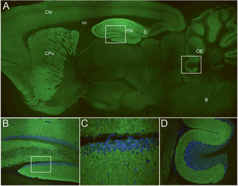
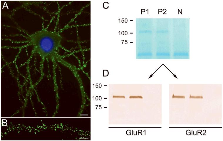
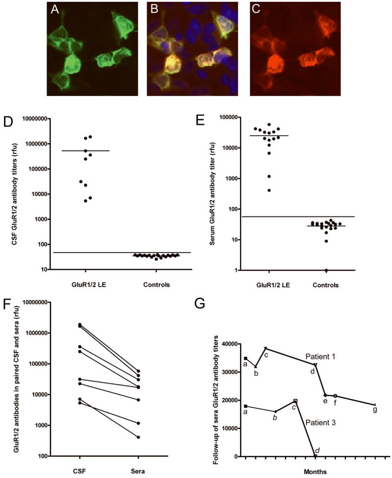
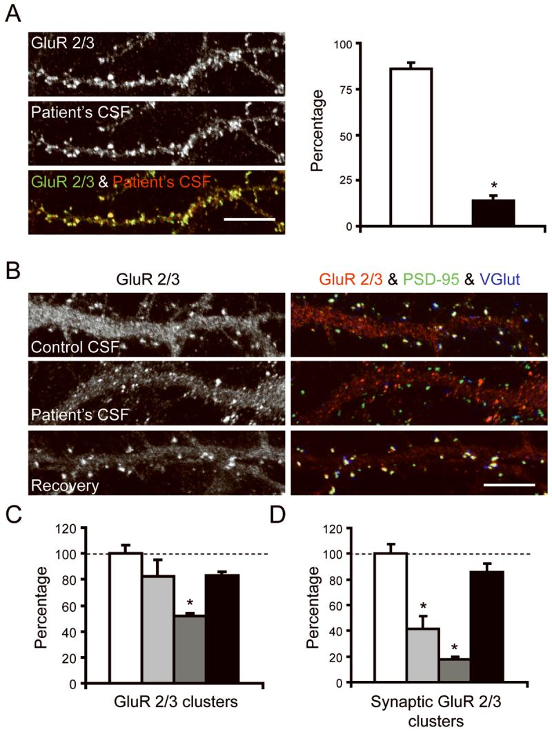
Methods: We conducted clinical analyses of 10 patients with LE. Immunoprecipitation and mass spectrometry were used to identify the antigens. Human embryonic kidney 293 cells expressing the antigens were used in immunocytochemistry and enzyme-linked immunoabsorption assay. The effect of patients' antibodies on cultures of live rat hippocampal neurons was determined with confocal microscopy.

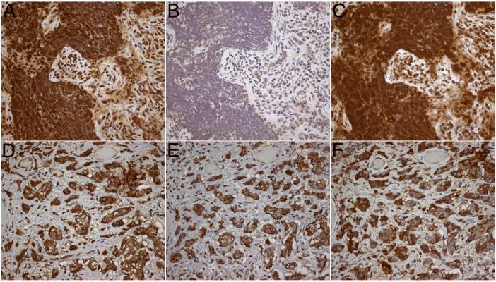
Results: Median age was 60 (38-87) years; 9 were women. Seven had tumors of the lung, breast, or thymus. Nine patients responded to immunotherapy or oncological therapy, but neurological relapses, without tumor recurrence, were frequent and influenced the long-term outcome. One untreated patient died of LE. All patients had antibodies against neuronal cell surface antigens that by immunoprecipitation were found to be the glutamate receptor 1 (GluR1) and GluR2 subunits of the alpha-amino-3-hydroxy-5-methyl-4-isoxazolepropionic acid receptor (AMPAR). Human embryonic kidney 293 cells expressing GluR1/2 reacted with all patients' sera or cerebrospinal fluid, providing a diagnostic test for the disorder. Application of antibodies to cultures of neurons significantly decreased the number of GluR2-containing AMPAR clusters at synapses with a smaller decrease in overall AMPAR cluster density; these effects were reversed after antibody removal.

Interpretation: Antibodies to GluR1/2 associate with LE that is often paraneoplastic, treatment responsive, and has a tendency to relapse. Our findings support an antibody-mediated pathogenesis in which patients' antibodies alter the synaptic localization and number of AMPARs.


Figures

Figure 1
Figure 1. Immunolabeling of rat brain by patients' antibodies
Sagittal section of rat brain incubated with the CSF of a patient with limbic encephalitis and novel antibodies. Note the intense reactivity of the patient's antibodies with the neuropil of hippocampus (Hip), subiculum (S), molecular layer of the cerebellum and Purkinje cells (CB), caudate-putamen (CPu), and cerebral cortex (Ctx). Other regions of the brain (e.g., corpus callosum (cc) and brainstem (B)) do not show significant immunolabeling. The areas boxed in A correspond to hippocampus and cerebellum and are shown at high magnification in B and D. The box in B is located at the dentate gyrus and is shown amplified in C. In panels B-D the nuclei of the cells is demonstrated with 4',6-diamidino-2-phenylindole (DAPI). Immunofluorescent technique, A × 2.5; B and D × 200; C × 400.
Figure 2
Figure 2. Patients antibodies react with extracellular epitopes and precipitate GluR1 and GluR2 subunits of the AMPAR
Culture of rat hipppocampal neurons incubated (live, non-permeabilized) with the CSF of a patient with LE. Note the intense reactivity of patient's antibodies with cell surface antigens (A); scale bar = 10 μm. Confocal microscopy suggests that the antigens are concentrated in puncta along dendrites (B); scale bar = 5 μm. The precipitation of these antigens using patients' antibodies is shown in a gel in which proteins are visualized with EZBlue (C). Note that two patients' antibodies (P1 and P2) precipitated antigen(s) that produce a single band at ~100 kDa; this band is not seen in the precipitate using CSF from a control individual (N). The band ~50 kDa corresponds to patients' IgG. Sequencing of the 100 kDa band using mass spectrometry demonstrated the GluR1 and GluR2 subunits of the AMPAR (not shown). Subsequent transfer of the gel to nitrocellulose and immunoblotting with antibodies specific for GluR1 and GluR2 confirmed that the 100 kDa band contained both GluR1 and GluR2 subunits (Panels in D).
Figure 3
Figure 3. Antibody reactivity with HEK293 cells co-transfected with GluR1/2 and quantitative ELISA studies
HEK293 cells co-transfected with GluR1/2 and immunostained with a patient's CSF (A) and an antibody specific for GluR2/3 (C). Note the co-localization of reactivities in (B). Protein extracts from HEK293 cells co-transfected with GluR1/2 were used to develop an ELISA (D-G). Panel D shows the titers of GluR1/2 antibodies in the CSF of 9 patients with GluR1/2 associated LE and 20 CSF randomly selected from controls; horizontal lines in each subgroup indicate the mean; the line across subgroups indicates 3 standard deviations above the mean (p < 0.001). Panel E shows the antibody titers of 8 patients' sera obtained during 15 episodes of LE (8 presentations and 7 relapses) and the titers of 20 controls (p < 0.001). Panel F compares the titers of GluR1/2 antibodies in paired CSF and serum samples in which the IgG has been normalized; note that in all 8 patients the antibody titers are higher in the CSF, indicating intrathecal synthesis of antibodies. Panel G shows the follow-up of serum antibody titers in two patients. Patient #1: a, titers at symptom presentation; b, 1 month after receiving 5-day treatment with intravenous methylprednisolone and plasma exchange (initially associated with substantial neurological improvement); c, first relapse of symptoms (after tapering corticosteroids), treated with intravenous methylprednisolone and IVIg; between c-d the patient had a second relapse partially treated with oral corticosteroids and anti-psychotic medication (she refused diagnostic tests and hospital admission); d, third relapse of symptoms, treated with IVIg and corticosteroids (partial neurological improvement); e-g, titers obtained while on azathioprine and stable neurological deficits. After the last time point (g) the patient was left with severe memory and behavioral deficits; for the last 3 years she has been living in a skill nursing facility with stable deficits (no follow-up titers available). Patient #3 had an episode of LE (that resolved spontaneously) 5 years before the current relapse; a, AMPAR antibody titers obtained at relapse of LE (which presented in association with anti-GAD related stiff-person syndrome); b-c, titers obtained during diagnostic studies and initial treatments (thymectomy, local radiation therapy); d, after intravenous corticosteroids (associated with dramatic improvement of LE). After the last time point (d), the patient remained only with symptoms of anti-GAD related stiff-person syndrome refractory to plasma exchange and IVIG; he eventually improved with chronic corticosteroids. (rfu = relative fluorescence, ELISA reader, Biotek Instruments, Winooski, Vermont).
Figure 4
Figure 4. Expression of GluR1 and GluR2 subunits by patients' tumors
The tumor of a patient with GluR1 antibodies demonstrates high level of expression of GluR1 (A), mild expression of GluR2 (B), and reactivity with patient's antibodies (C). The tumor of a patient with GluR2 antibodies demonstrates high level of expression of GluR1 (D) and GluR2 (E), and reactivity with patient's antibodies (F). Tissues immunostained with the indicated avidin-biotin peroxidase method, and mildly counterstained with hematoxylin. All panels x400.
Figure 5
Figure 5. Patient's antibodies selectively bind to GluR2 and alter the number and localization of AMPAR in live neurons
Panel A shows 17 div hippocampal neurons immunostained with patient's CSF (b+w, red) and with a commercial antibody specific for GluR 2/3 (b+w, green). White column indicates the number of GluR2/3-containing clusters labeled by patient's CSF; black column indicates the number of GluR2/3-containing clusters that are not labeled by patient's CSF. Since patient's antibodies react with GluR2, but not GluR3 (see text), the findings indicate that nearly all clusters labeled with patient's CSF correspond to GluR2 (91%, yellow puncta in overlay). Scale bar = 5 μm. Panels B-D show hippocampal neurons cultured with control CSF or patient's CSF and subsequently immunostained for GluR 2/3 (b+w, red), the postsynaptic marker PSD-95 (green), and the pre-synaptic marker VGlut (blue). White columns indicate the number of GluR2/3-containing clusters in neurons cultured for 6 days with control CSF; Light grey columns indicate the number of clusters in neurons cultured for 3 days with patient's CSF; Dark grey columns indicates the number of clusters in neurons cultured for 6 days with patient's CSF; Black columns indicate the number of clusters in neurons cultured for 3 days with patient's CSF and subsequently cultured for 3 days with control CSF (3 day recovery). Note that patient's CSF, applied from 11-17 div (6 day treatment), reduces the number of GluR 2/3 labeled puncta compared with cultures exposed to control CSF (Panel B “GluR 2/3” and panel C; p < 0.001). Moreover, the patient's CSF applied for 3 or 6 days, but not the control CSF, reduces the number of GluR 2/3 clusters that colocalize with PSD-95 (yellow puncta = postsynaptic AMPAR), and the number of GluR 2/3 clusters that colocalize with VGlut (white puncta = presynaptic AMPAR) (Panel B “GluR 2/3 & PSD-95 & VGlut” and panel D; p < 0.01). These effects were reversed after removing the antibodies from the cultures and allowing the neurons to recover for 3 days (panels C and D). Scale bar = 5 μm. For each condition, a minimum of 6-15 neurons was examined on each of 2-3 coverslips in 3 independent experiments. Neurons were randomly selected for analysis. The number of synaptic clusters was calculated per 20 μm and expressed as a percentage of control values. Results were analyzed using the Kruskal-Wallis non-parametric ANOVA followed by Dunn's pairwise comparison.

References

    1. Corsellis JA, Goldberg GJ, Norton AR. “Limbic encephalitis” and its association with carcinoma. Brain. 1968;91:481–496. - PubMed
    1. Gultekin SH, Rosenfeld MR, Voltz R, Eichen J, Posner JB, Dalmau J. Paraneoplastic limbic encephalitis: neurological symptoms, immunological findings and tumour association in 50 patients. Brain. 2000;123(Pt 7):1481–1494. - PubMed
    1. Bataller L, Kleopa KA, Wu GF, Rossi JE, Rosenfeld MR, Dalmau J. Autoimmune limbic encephalitis in 39 patients: immunophenotypes and outcomes. J Neurol Neurosurg Psychiatry. 2007;78:381–385. - PMC - PubMed
    1. Ances BM, Vitaliani R, Taylor RA, et al. Treatment-responsive limbic encephalitis identified by neuropil antibodies: MRI and PET correlates. Brain. 2005;128:1764–1777. - PMC - PubMed
    1. Graus F, Saiz A, Lai M, et al. Neuronal surface antigen antibodies in limbic encephalitis: clinical-immunologic associations. Neurology. 2008;71:930–936. - PMC - PubMed

Publication types

MeSH terms

LinkOut - more resources