. 2009 Oct;15(10):3099-108.
doi: 10.1089/ten.TEA.2008.0502.

Cell-induced alignment augments twitch force in fibrin gel-based engineered myocardium via gap junction modification

Affiliations

Cell-induced alignment augments twitch force in fibrin gel-based engineered myocardium via gap junction modification

Lauren D Black 3rd et al. Tissue Eng Part A. 2009 Oct.

Abstract

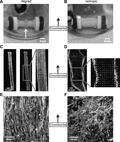
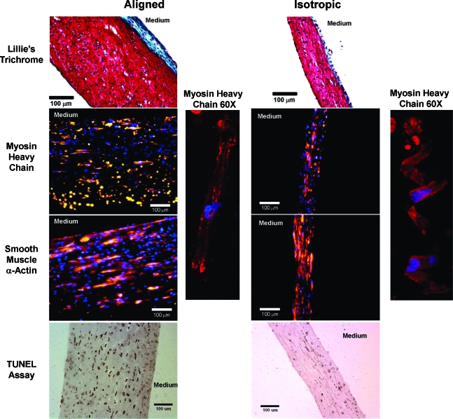
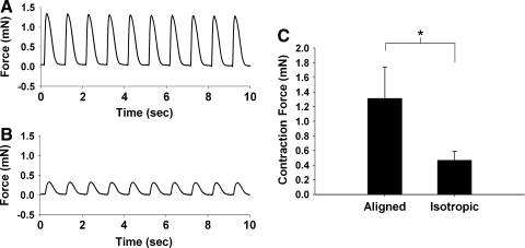
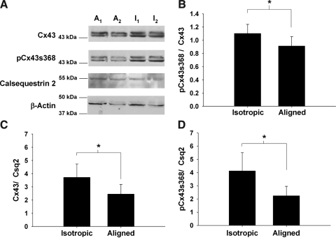
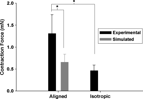
A high-potential therapy for repairing the heart post-myocardial infarction is the implantation of tissue-engineered myocardium. While several groups have developed constructs that mimic the aligned structure of the native myocardium, to date no one has investigated the particular functional benefits conferred by alignment. In this study we created myocardial constructs in both aligned and isotropic configurations by entrapping neonatal rat cardiac cells in fibrin gel. Constructs were cultured statically for 2 weeks, and then characterized. Histological staining showed spread cells that express typical cardiac cell markers in both configurations. Isotropic constructs had higher final cell and collagen densities, but lower passive mechanical properties than aligned constructs. Twitch force associated with electrical pacing, however, was 181% higher in aligned constructs, and this improvement was greater than what would be expected from merely aligning the cells in the isotropic constructs in the force measurement direction. Our hypothesis was that this was due to improved gap junction formation/function facilitated by cell alignment, and further analyses of the twitch force data, as well as Western blot results of connexin 43 expression and phosphorylation state, support this hypothesis. Regardless of the specific mechanism, the results presented in this study underscore the importance of recapitulating the anisotropy of the native tissue in engineered myocardium.


Figures

FIG. 1.
FIG. 1.
Images of isotropic (A) and aligned (B) constructs after 14 days in culture with corresponding polarized light alignment images (C, D). In (C, D) the constructs have been cut axially and splayed flat. The grayscale values represent the birefringence of the tissue, a measure of the strength of alignment, and the lines (black in C, white in D) indicate the local average direction of alignment in the tissue. Note that the aligned construct (D) is highly aligned in the circumferential (vertical) direction, while the isotropic construct (C) does not possess alignment. Cellular alignment, measured via staining of filamentous actin, matched matrix alignment assessed via polarized light for both construct types (E, F).
FIG. 2.
FIG. 2.
Representative histological sections imaged at 10 × for aligned and isotropic constructs. Constructs were stained with Lillie's Trichrome stain for matrix components (top row), and antibodies for myosin heavy chain (red stain, second row) and α-smooth muscle actin (red, third row). Note the inset of myosin heavy chain staining at 60× showing myosin fiber development and cross-striation typical of native myocytes (right of second and third rows). In addition, a TUNEL assay was conducted to assess apoptotic cells in aligned and isotropic constructs (bottom row). Note that TUNEL-positive nuclei stain dark brown or black to indicate apoptotic cells. Blue represents a DAPI counterstain for cell nuclei in the middle two rows. The surface in contact with the culture medium is noted for each image. In addition, the blue-green color of collagen stained in the trichrome stain is labeled in these images for both construct types. Color images available online at www.liebertonline.com/ten.
FIG. 3.
FIG. 3.
(A) Final cell number in millions for aligned and isotropic constructs after 14 days in culture. Note that the isotropic constructs have almost twice as many cells remaining after 14 days. (B) Passive mechanical properties (modulus and UTS) for aligned and isotropic constructs after 14 days in culture. Total protein density (C) and collagen density (D) after 14 days in culture for aligned and isotropic constructs. Note that the isotropic constructs have higher total protein and collagen densities than the aligned constructs. *p < 0.05 (n = 14 for all groups). UTS, ultimate tensile strength.
FIG. 4.
FIG. 4.
Representative force traces during 1 Hz pacing versus time plots for an aligned (A) and an isotropic (B) construct. (C) Pooled twitch force data for 14-day-old aligned (left) and isotropic (right) constructs at 10 mN preload during 1 Hz pacing. *p < 0.05 (n = 14 for both groups).
FIG. 5.
FIG. 5.
(A) Representative Western blots for connexin 43 (Cx43), connexin 43 phosphorylated at serine 368 (pCx43s368), calsequestrin 2 (Csq2), and β-actin for two aligned (A1, A2) and two isotropic (I1, I2) constructs. (B) The ratio of pCx43s368 expression to Cx43 expression for aligned and isotropic constructs. (C) The ratio of Cx43 to Csq2 for aligned and isotropic constructs. (D) The ratio of pCx43s368 expression to Csq2 expression for aligned and isotropic constructs. *p < 0.05 (n = 6 for all groups).
FIG. 6.
FIG. 6.
Twitch force data for experimental data from aligned and isotropic constructs as well as aligned constructs simulated from isotropic data (see text). *p < 0.05 (n = 14 for all groups).

References

    1. Hoyert D.L. Heron M.P. Murphy S.L. Kung H.C. Deaths: final data for 2003. Natl Vital Stat Rep. 2006;54:1. - PubMed
    1. Miniati D.N. Robbins R.C. Heart transplantation: a thirty-year perspective. Annu Rev Med. 2002;53:189. - PubMed
    1. Bursac N. Papadaki M. Cohen R.J. Schoen F.J. Eisenberg S.R. Carrier R. Vunjak-Novakovic G. Freed L.E. Cardiac muscle tissue engineering: toward an in vitro model for electrophysiological studies. Am J Physiol. 1999;277:H433. - PubMed
    1. Leor J. Aboulafia-Etzion S. Dar A. Shapiro L. Barbash I.M. Battler A. Granot Y. Cohen S. Bioengineered cardiac grafts: a new approach to repair the infarcted myocardium? Circulation. 2000;102:III56. - PubMed
    1. Radisic M. Park H. Shing H. Consi T. Schoen F.J. Langer R. Freed L.E. Vunjak-Novakovic G. Functional assembly of engineered myocardium by electrical stimulation of cardiac myocytes cultured on scaffolds. Proc Natl Acad Sci USA. 2004;101:18129. - PMC - PubMed

Publication types

LinkOut - more resources