Review
. 2009 Mar:1156:260-93.
doi: 10.1111/j.1749-6632.2009.04420.x.

What's new in neuroimaging methods?

Affiliations
Review

What's new in neuroimaging methods?

Peter A Bandettini. Ann N Y Acad Sci. 2009 Mar.

Abstract

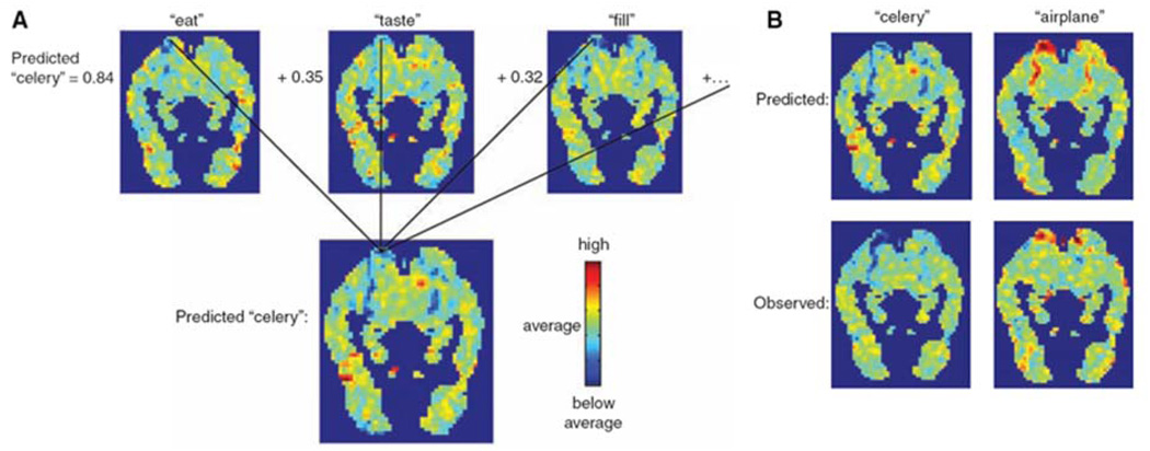
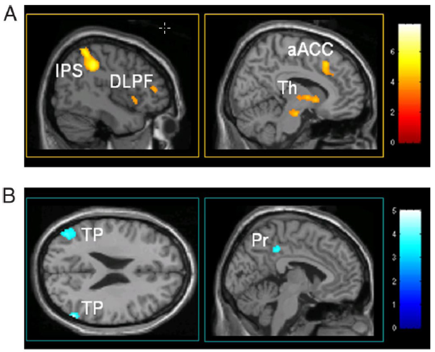
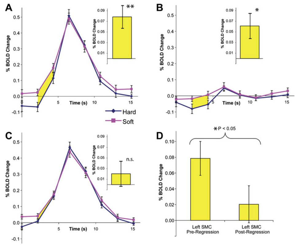
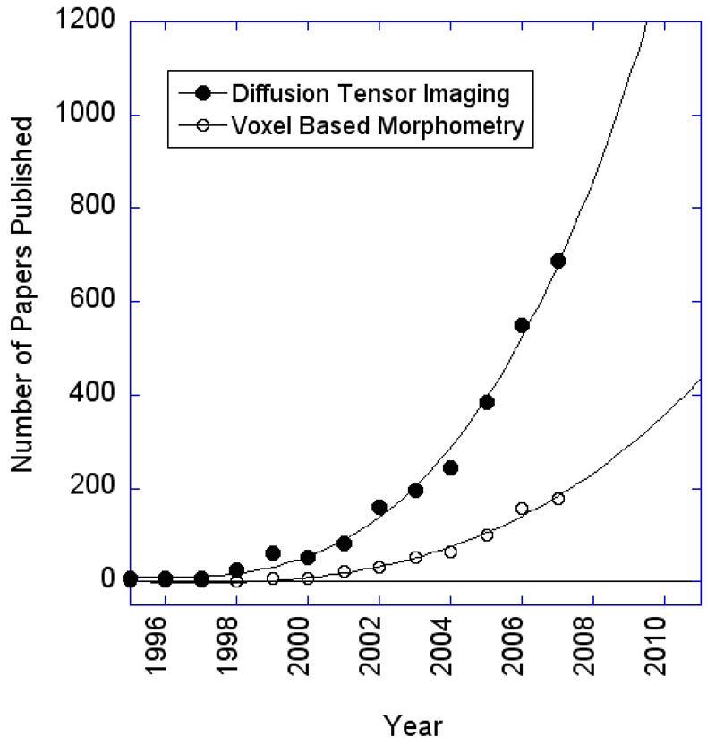
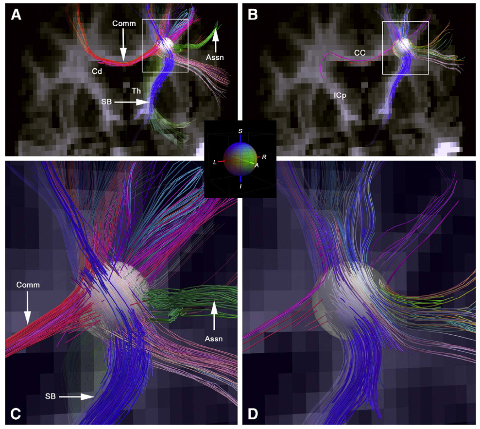
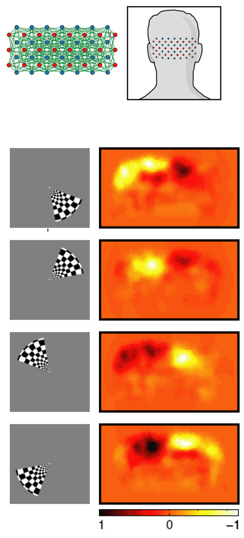
The rapid advancement of neuroimaging methodology and its growing availability has transformed neuroscience research. The answers to many questions that we ask about how the brain is organized depend on the quality of data that we are able to obtain about the locations, dynamics, fluctuations, magnitudes, and types of brain activity and structural changes. In this review an attempt is made to take a snapshot of the cutting edge of a small component of the very rapidly evolving field of neuroimaging. For each area covered, a brief context is provided along with a summary of a few of the current developments and issues. Then, several outstanding papers, published in the past year or so, are described, providing an example of the directions in which each area is progressing. The areas covered include functional magnetic resonance imaging (fMRI), voxel-based morphometry (VBM), diffusion tensor imaging (DTI), electroencephalography (EEG), magnetoencephalography (MEG), optical imaging, and positron emission tomography (PET). More detail is included on fMRI; its subsections include fMRI interpretation, new fMRI contrasts, MRI technology, MRI paradigms and processing, and endogenous oscillations in fMRI.


Figures

Figure 1
Figure 1
Bar chart of the number of abstracts for each of the 12 categories in Neuroimaging presented at the 2007 Organization for Human Brain Mapping meeting. The focus of this current review article is on neuroimaging methods which mostly covers “modeling and analysis,” “ imaging techniques and contrast mechanisms,” and some “physiology metabolism and neurotransmission.” Overall, the topics covered in this article consist of, at most, about a third of the field of neuroimaging.
Figure 2
Figure 2
Obtained from (Muthukumaraswamy & Singh, 2008). This shows the temporal frequency tuning curves for peak responses in fMRI and MEG Gamma frequency (40×60Hz) data in primary visual cortex, clearly indicating a difference between fMRI and MEG responses. No spatial frequency dependence is seen in fMRI, while with MEG, as strong spatial frequency dependence is demonstrated. In addition, with MEG, the tuning curves are show a more flat responsivity than the fMRI curves.
Figure 3
Figure 3
Obtained from (Sun et al., 2007). This is NOT an ocular dominance column map. It is an fMRI-derived temporal frequency domain map, showing the fine structure of areas in primary visual cortex that are either selective to high temporal frequency visual stimuli (blue) or low temporal frequency visual stimuli (yellow). This is the first demonstration of spatial organization of cortex based on temporal frequency.
Figure 4
Figure 4
Plot of the highest current MRI field strength used for human imaging as it has increased over the years, showing a surprising linear trend.
Figure 5
Figure 5
Pie chart showing the current distribution (as of the summer of 2008), by country, of 7 Tesla human scanners. (Data is courtesy of Hellmut Merkle, NINDS)
Figure 6
Figure 6
Obtained from (Duyn et al., 2007). This is an illustration of the image quality of magnitude and phase images obtained at 7T. The GRE data, left and center, had a resolution of 240 × 240 um with a scan duration of 6.5 min, whereas the MP-RAGE data on the right had a resolution of 480 × 480 um and a scan time of 20 min. The scale bar shows the frequency shifts corresponding to the phase changes.
Figure 7
Figure 7
Obtained from (Yacoub et al., 2008). This shows ocular dominance columns for two subjects in panels a and b. Red and blue represent voxels that showed preference to right and left eye stimulation, respectively. Maps shown in panels c and d, show maps in the same cortical areas, of orientation preference. The black and white circles on the orientation preference maps show where multiple preferences converge. These form “pinwheel center.” White circles indicate clockwise pinwheels and black circles indicate counterclockwise pinwheels. The white bars at the base of the images are 0.5 mm in length.
Figure 8
Figure 8
Obtained from (Kay et al., 2008). This is a schematic diagram showing the steps in the fMRI decoding experiment. In the first stage, fMRI data were recorded as subjects viewed a large collection of natural images. From these data, receptive field models for each voxel were created, based on a Gabor wavelet pyramid. These describe tuning along the dimensions of space, orientation, and spatial frequency. In the next stage, fMRI data were recorded while each subject viewed a collection of novel natural images. For each brain map, an attempt is made to identify the image that was seen using the receptive field models.
Figure 9
Figure 9
Obtained from (Mitchell et al., 2008). This figure shows the process by which predicted fMRI maps were created for specific stimulus words. The top three maps in A. are learned spatial coefficients for 3 of the 25 semantic features related to “celery” (“eat,” “taste,” and “fill”). The co-occurrence in normal speech of each of these features is shown on the left of the images. These weighted images are summed to create the predicted image. In B. are two examples of predicted maps (above), showing a clear difference between the two, with observed maps (below) showing a relatively high level of similarity between predicted and observed.
Figure 10
Figure 10
Obtained from (Hasson et al., 2008). This shows a summary of their clever experiment testing which cortical areas are sensitive and insensitive to time reversals in movie sequences. Plots are from area MT+. A. Representative frames from the silent films used. Average time courses for B. two forward, and C. two backward presentations. D. Superimposed plots of a time-reversed version of the backwards movie time course and the forward time course (both time course shifted appropriately to account for the hemodynamic delay). E. Cross correlations of various iterations of the two forward time courses with themselves (black), backward with themselves (red), reversed backward with forward (green), and backward with forward (in blue - as expected showing minimal correlation) showing that at least in MT+, temporal order is certainly not critical for activation.
Figure 11
Figure 11
Obtained from (Boly et al., 2008). Maps of baseline activity that predicts conscious perception of subsequent somatosensory stimuli. A. Increased activity of the medial thalamus (Th), dorsolateral prefrontal cortex (DLPF), intraparietal sulcus/posterior parietal cortex (IPS), and anterior cingulate cortex (aACC) 3 sec before stimulus presentation predicts perception of low-intensity sensory stimuli. B. Decreased baseline activity in the default brain network - posterior cingulate/precuneus (Pr), bilateral temporal/parietal junctions (TP) - exerts an enhancing effect on perception of subsequent somatosensory stimuli.
Figure 12
Figure 12
Obtained from (Fox et al., 2007). This is a demonstration that coherent spontaneous activity in the motor cortex influences the force at which subjects press a button. A. Average left SMC BOLD time courses for the hard (blue) and soft (red) button presses. Yellow indicates significant differences between them. The same time courses for the right SMC also shows a BOLD-behavior effect. C. After spontaneous fluctuations were regressed out, the effect disappears. D. Summary of the comparison of A and C.
Figure 13
Figure 13
Obtained from (Ilg et al., 2008). This shows practice related changes in activation and in gray matter density related to performing the task of mirror reading. The green areas indicate the overall activation. The blue area is where the activation induced signal change increased with practice. The red area is where the activation induced signal change decreased with practice. Lastly, the white area is where the change in gray matter density was found.
Figure 14
Figure 14
Results of a literature search using “Scopus” of all diffusion tensor imaging and voxel based morphometry imaging studies published in the past 12 years. A rapid growth in both is seen after 2002.
Figure 15
Figure 15
Obtained from (Wedeen et al., 2008). These panels show crossing fibers within the centrum semiovale of in vivo human brain using DSI (A, C - magnification of A) and DTI (B, D -magnification of B). Assn, long association fibers; CC corpus callosum; Cd caudate nucleus; Comm, commissural fibers; ICp, posterior limb of internal capsule; SB subcortical bundle projection fibers; Th, thalamus. DSI shows superior fiber tracking resolution over DTI.
Figure 16
Figure 16
Obtained from (Zeff et al., 2007). This shows polar angle retinotopic mapping with diffuse optical tomography.

References

    1. Aguirre GK, Detre JA, Zarahn E, Alsop DC. Experimental design and the relative sensitivity of BOLD and perfusion fMRI. Neuroimage. 2002;15(3):488–500. - PubMed
    1. Ances BM, Leontiev O, Perthen JE, Liang C, Lansing AE, Buxton RB. Regional differences in the coupling of cerebral blood flow and oxygen metabolism changes in response to activation: Implications for BOLD-fMRI. NeuroImage. 2008;39(4):1510–1521. - PMC - PubMed
    1. Apkarian AV, Sosa Y, Sonty S, Levy RM, Harden RN, Parrish TB, et al. Chronic back pain is associated with decreased prefrontal and thalamic gray matter density. Journal of Neuroscience. 2004;24(46):10410–10415. - PMC - PubMed
    1. Ashburner J, Friston KJ. Voxel-based morphometry-The methods. Neuroimage. 2000;11(6 I):805–821. - PubMed
    1. Baliki MN, Geha PY, Apkarian AV, Chialvo DR. Beyond feeling: Chronic pain hurts the brain, disrupting the default-mode network dynamics. Journal of Neuroscience. 2008;28(6):1398–1403. - PMC - PubMed

MeSH terms