

Extracorporeal shock wave lithotripsy at 60 shock waves/min reduces renal injury in a porcine model
- PMID: 19338532
- PMCID: PMC2888935
- DOI: 10.1111/j.1464-410X.2009.08520.x
Extracorporeal shock wave lithotripsy at 60 shock waves/min reduces renal injury in a porcine model
Abstract
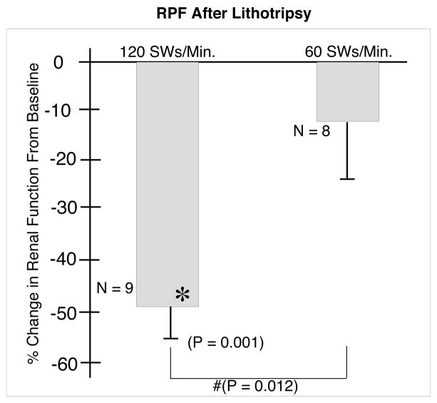
Objective: To determine if extracorporeal shock wave lithotripsy (ESWL) at 60 shock waves (SWs)/min reduces renal damage and haemodynamic impairment compared to treatment at 120 SWs/min.
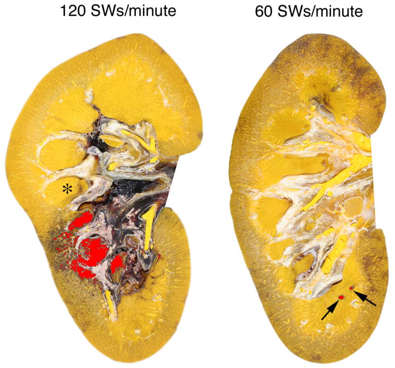
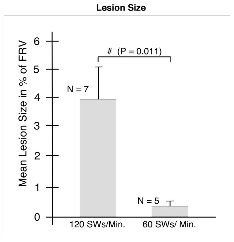
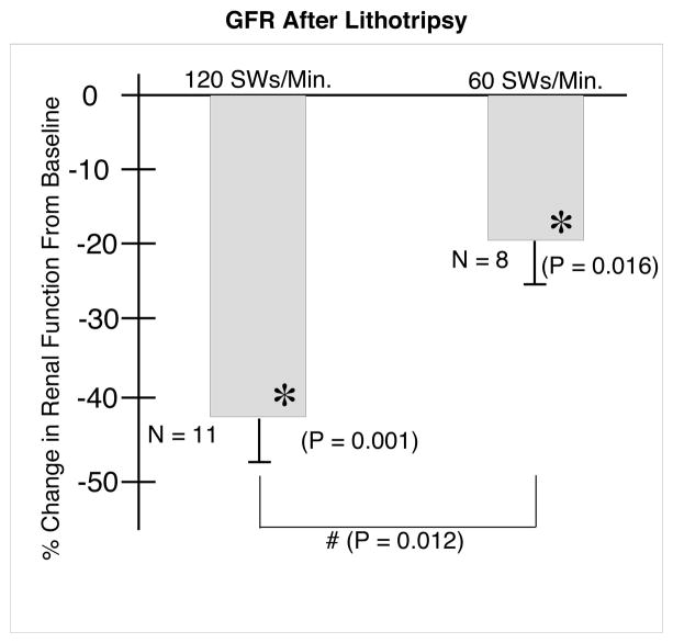
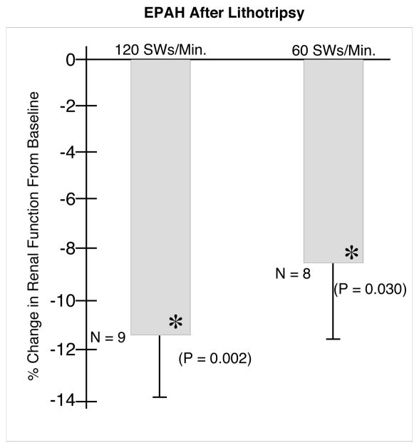
Materials and methods: One kidney in each of 19 juvenile pigs (7-8 weeks old) was treated at 120 or at 60 SWs/min (2000 SWs, 24 kV) with an unmodified HM-3 lithotripter (Dornier Medical Systems, Kennesaw, GA, USA). Renal function was determined before and after ESWL treatment by inulin clearance, extraction and clearance of para-aminohippuric acid. Both kidneys were then removed to measure parenchymal lesion size by sectioning the entire kidney and quantifying the size of the haemorrhagic lesion in each slice.
Results: ESWL at 60 SWs/min significantly reduced the size of the acute morphological lesion compared to 120 SWs/min (0.42% vs 3.93% of functional renal volume, P = 0.011) and blunted the decrease in glomerular filtration rate and renal plasma flow normally seen after treatment at 120 SWs/min.
Conclusions: Treatment at a firing rate of 60 SWs/min produces less morphological injury and causes less alteration in renal haemodynamics than treatment at 120 SWs/min in the pig model of ESWL-induced renal injury.
Figures
References
-
- Evan AP, Willis LR, Connors BA, McAteer JA, Lingeman JE. Renal injury by extracorporeal shock wave lithotripsy. J Endourol. 1991;5:25–35.
-
- Kaude JV, Williams CM, Millner MR, Scott JN, Finlayson B. Renal morphology and function immediately after extracorporeal shock wave lithotripsy. Am J Roentgenol. 1985;145:305–13. - PubMed
-
- Orozc Fainas R, Iglesias Prieto JI, Massarrah Halabi J, Mancebo Gomez JM, Perez-Castro Ellendt E. Renal hematoma after extracorporeal shockwave lithotripsy in a series of 324 consecutive sessions with the Doli-S lithotripter: incidents, characteristics, multifactorial analysis and review. Arch Esp Urol. 2008;61:889–914. - PubMed
-
- Krishnamurthi V, Streem SB. Long-term radiographic and functional outcome of extracorporeal shock wave lithotripsy induced perirenal hematomas. J Urol. 1995;154:1673–5. - PubMed
Publication types
MeSH terms
Grants and funding
LinkOut - more resources
Full Text Sources
