

TDP-43 redistribution is an early event in sporadic amyotrophic lateral sclerosis
- PMID: 19338576
- PMCID: PMC8094784
- DOI: 10.1111/j.1750-3639.2009.00284.x
TDP-43 redistribution is an early event in sporadic amyotrophic lateral sclerosis
Abstract
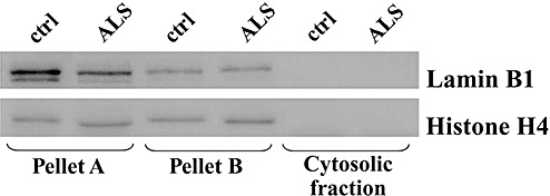
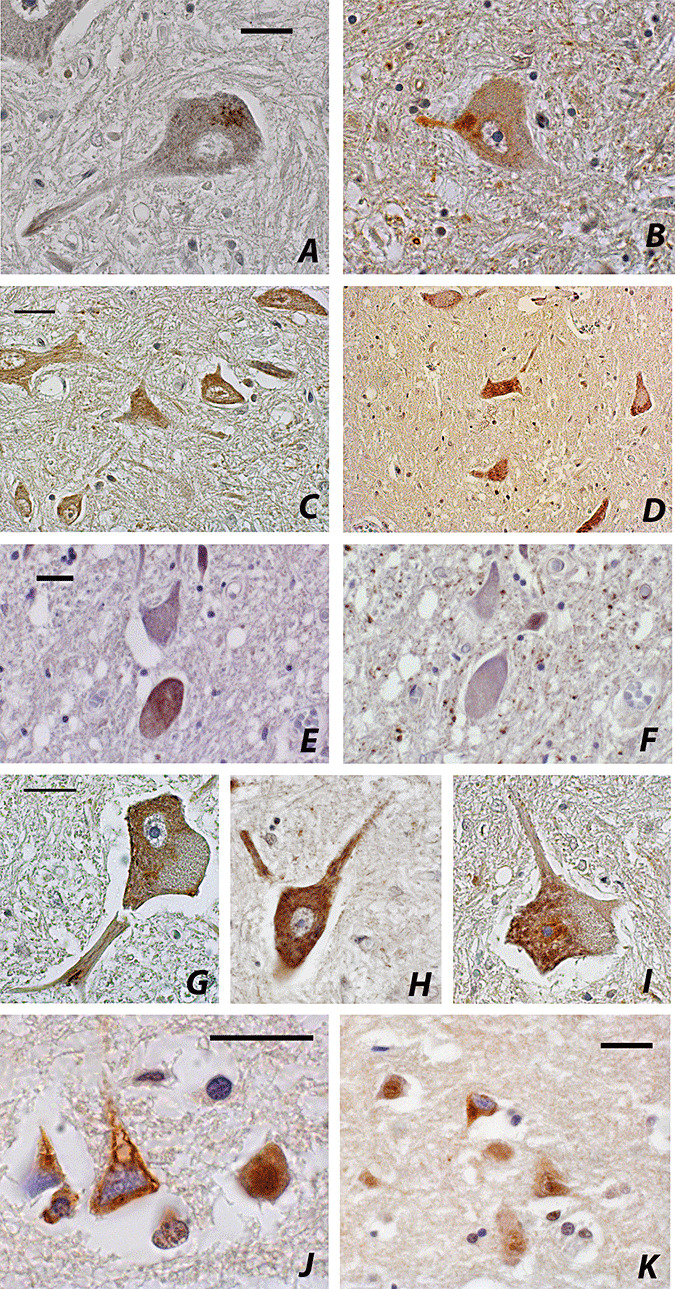
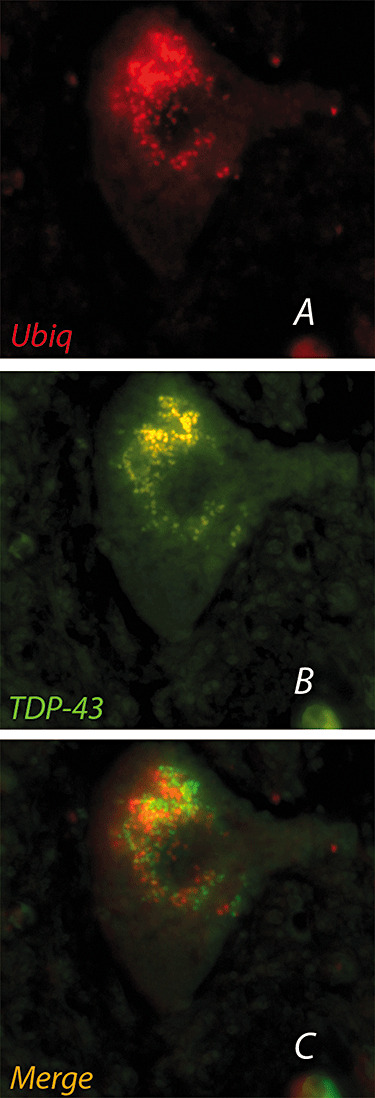
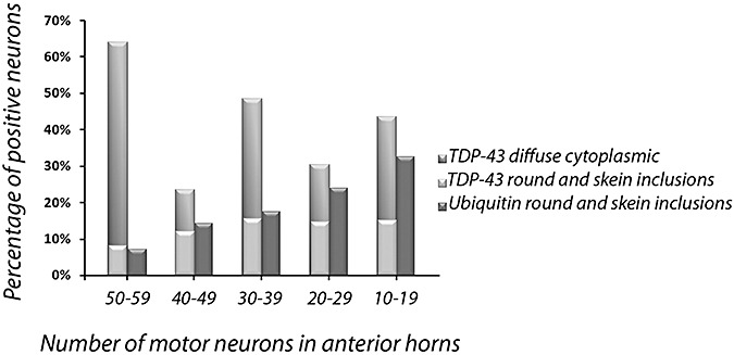
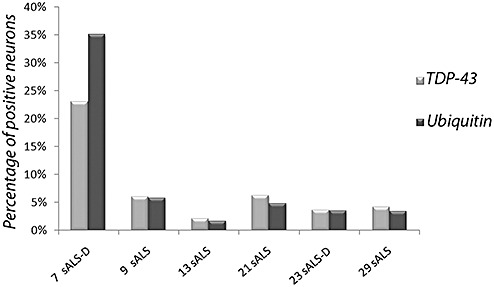
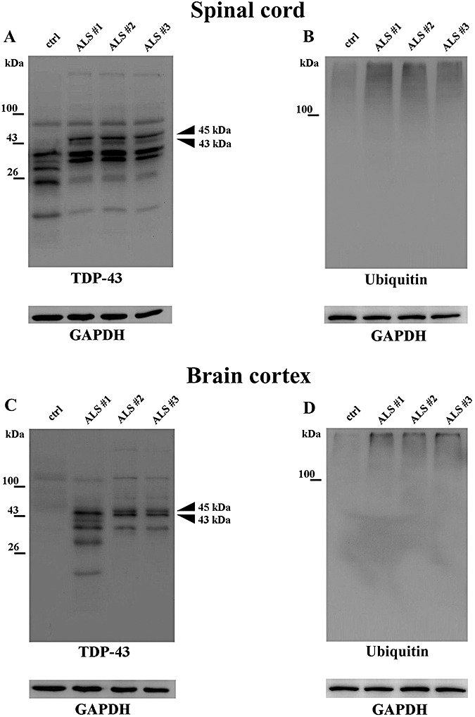
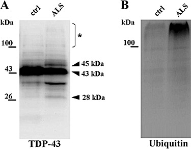
Amyotrophic lateral sclerosis (ALS) is a neurodegenerative disorder consisting of progressive loss of motor neurons. TDP-43 has been identified as a component of ubiquitin-immunoreactive inclusions of motor neurons in ALS. We focused on the diffuse cytoplasmic TDP-43 immunoreactivity in ALS neurons, and quantitatively assessed it in comparison with skein/round TDP-43 and ubiquitin immunostaining in motor neurons of 30 sporadic ALS cases. The percentage of spinal motor neurons with cytoplasmic TDP-43 immunoreactivity was higher than that of ubiquitin-immunoreactive ones. The percentage of TDP-43-positive motor neurons was independent of neuron counts in anterior horns, while the percentage of ubiquitinated neurons was inversely correlated. Aiming to define the cytosolic localization of TDP-43, the immunoblot analysis of spinal cord and frontal cortex showed that full-length TDP-43, the 45 kDa form and ubiquitinated TDP-43 are found in the soluble inclusion-free fraction. The present data suggest that delocalization, accumulation and ubiquitination of TDP-43 in the cytoplasm of motor neurons are early dysfunctions in the cascade of the events leading to motor neuron degeneration in ALS, preceding the formation of insoluble inclusion bodies. Being cytoplasmic accumulation an ongoing event during the course of the illness, a therapeutic approach to this incurable disease can be envisaged.
Figures
References
-
- Arai T, Hasegawa M, Akiyama H, Ikeda K, Nonaka T, Mori H et al (2006) TDP‐43 is a component of ubiquitin‐positive inclusions in frontotemporal lobar degeneration and amyotrophic lateral sclerosis. Biochem Biophys Res Commun 351:602–611. - PubMed
-
- Bjerrum OJ, Schafer‐Nielsen C (1986) Buffer system and transfer parameters for semi‐dry electroblotting with a horizontal apparatus. In: Electrophoresis'68. Dunn MJ (ed.), pp. 315–327. VCH: Weinheim.
-
- Brandmeir NJ, Geser F, Kwong LK, Zimmerman E, Qian J, Lee VM, Trojanowski JQ (2007) Severe subcortical TDP‐43 pathology in sporadic frontotemporal lobar degeneration with motor neuron disease. Acta Neuropathol 115:147–149. - PubMed
-
- Brooks BR, Miller RG, Swash M, Munsat TL, World Federation of Neurology Research Group on Motor Neuron Diseases (2000) El Escorial revisited: revised criteria for the diagnosis of amyotrophic lateral sclerosis. Amyotroph Lateral Scler Other Motor Neuron Disord 1:293–299. - PubMed
Publication types
MeSH terms
Substances
LinkOut - more resources
Full Text Sources
Other Literature Sources
Medical
Miscellaneous
