

Structure and mechanical properties of Ank/Ank mutant mouse dental tissues--an animal model for studying periodontal regeneration
- PMID: 19338977
- PMCID: PMC5988219
- DOI: 10.1016/j.archoralbio.2009.02.011
Structure and mechanical properties of Ank/Ank mutant mouse dental tissues--an animal model for studying periodontal regeneration
Abstract
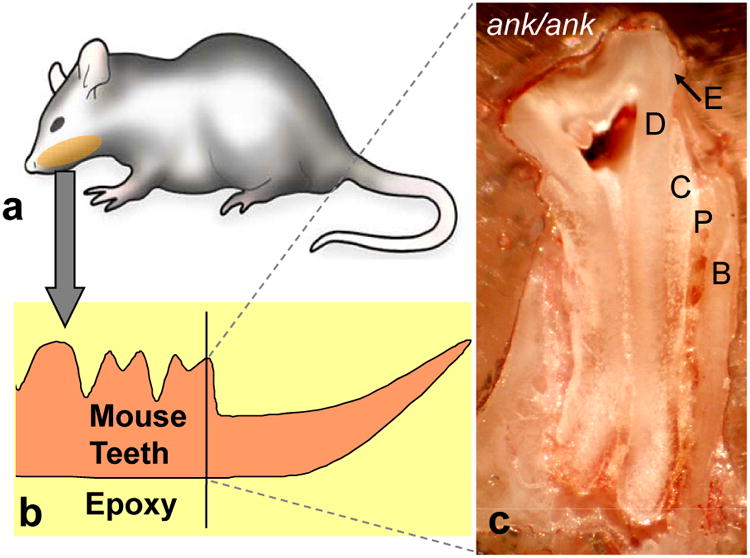
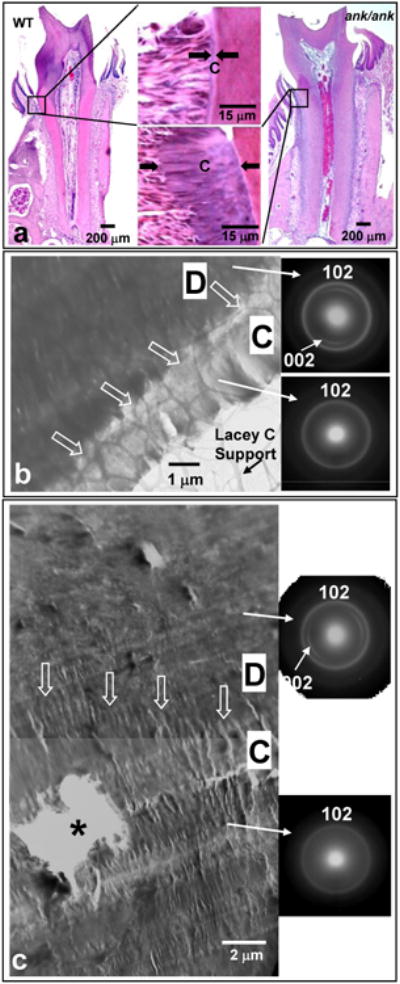
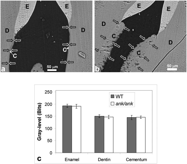
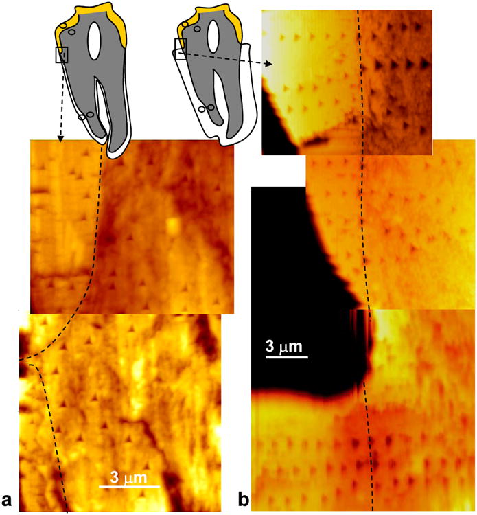
Enamel, dentine and cementum are dental tissues with distinct functional properties associated with their unique hierarchical structures. Some potential ways to repair or regenerate lost tooth structures have been revealed in our studies focused on examining teeth obtained from mice with mutations at the mouse progressive ankylosis (ank) locus. Previous studies have shown that mice with such mutations have decreased levels of extracellular inorganic pyrophosphate (PP(i)) at local sites resulting in ectopic calcification in joint areas and in formation of a significantly thicker cementum layer when compared with age-matched wild-type (WT) tissue [Ho AM, Johnson MD, Kingsley DM. Role of the mouse ank gene in control of tissue calcification and arthritis. Science 2000;289:265-70; Nociti Jr FH, Berry JE, Foster BL, Gurley KA, Kingsley DM, Takata T, et al. Cementum: a phosphate-sensitive tissue. J Dent Res 2002;81:817-21]. As a next step, to determine the quality of the cementum tissue formed in mice with a mutation in the ank gene (ank/ank), we compared the microstructure and mechanical properties of cementum and other dental tissues in mature ank/ank vs. age-matched WT mice. Backscattered scanning electron microscopy (SEM) imaging and transmission electron microscopy (TEM) analyses on mineralized tissues revealed no decrease in the extent of mineralization between ank/ank cementum vs. WT controls. Atomic-force-microscopy-based nanoindentation performed on enamel, dentine or cementum of ank/ank vs. age-matched WT molars revealed no significant difference in any of the tested tissues in terms of hardness and elastic modulus. These results indicate that the tissue quality was not compromised in ank/ank mice despite faster rate of formation and more abundant cementum when compared with age-matched WT mice. In conclusion, these data suggest that this animal model can be utilized for studies focused on defining mechanisms to promote cementum formation without loss of mechanical integrity.
Figures
References
-
- Ho AM, Johnson MD, Kingsley DM. Role of the mouse ank gene in control of tissue calcification and arthritis. Science. 2000;289:265–270. - PubMed
-
- Nociti FH, Jr, Berry JE, Foster BL, Gurley KA, Kingsley DM, Takata T, Miyauchi M, Somerman MJ. Cementum: a phosphate-sensitive tissue. Journal of Dental Research. 2002;81:817–821. - PubMed
-
- Polimeni G, Xiropaidis AV, Wikesjo UM. Biology and principles of periodontal wound healing/regeneration. Periodontol. 2006;200041:30–47. - PubMed
-
- Abukawa H, Papadaki M, Abulikemu M, Leaf J, Vacanti JP, Kaban LB, Troulis MJ. The engineering of craniofacial tissues in the laboratory: a review of biomaterials for scaffolds and implant coatings. Dent Clin North Am. 2006;50:205–16. - PubMed
Publication types
MeSH terms
Substances
Grants and funding
LinkOut - more resources
Full Text Sources
Other Literature Sources
Molecular Biology Databases
Research Materials
Miscellaneous
