Credentialing a preclinical mouse model of alveolar rhabdomyosarcoma
- PMID: 19339268
- PMCID: PMC2789740
- DOI: 10.1158/0008-5472.CAN-08-3723
Credentialing a preclinical mouse model of alveolar rhabdomyosarcoma
Abstract
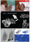
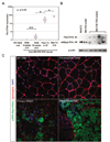
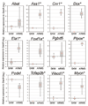
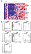
The highly aggressive muscle cancer alveolar rhabdomyosarcoma (ARMS) is one of the most common soft tissue sarcoma of childhood, yet the outcome for the unresectable and metastatic disease is dismal and unchanged for nearly three decades. To better understand the pathogenesis of this disease and to facilitate novel preclinical approaches, we previously developed a conditional mouse model of ARMS by faithfully recapitulating the genetic mutations observed in the human disease, i.e., activation of Pax3:Fkhr fusion gene with either p53 or Cdkn2a inactivation. In this report, we show that this model recapitulates the immunohistochemical profile and the rapid progression of the human disease. We show that Pax3:Fkhr expression increases during late preneoplasia but tumor cells undergoing metastasis are under apparent selection for Pax3:Fkhr expression. At a whole-genome level, a cross-species gene set enrichment analysis and metagene projection study showed that our mouse model is most similar to human ARMS when compared with other pediatric cancers. We have defined an expression profile conserved between mouse and human ARMS, as well as a Pax3:Fkhr signature, including the target gene, SKP2. We further identified 7 "druggable" kinases overexpressed across species. The data affirm the accuracy of this genetically engineered mouse model.
Conflict of interest statement
C.K. is co-founder of Numira Biosciences, which has licensed micro-CT-based Virtual Histology from UTHSCSA.
Figures


Similar articles
-
Structural and functional studies of FKHR-PAX3, a reciprocal fusion gene of the t(2;13) chromosomal translocation in alveolar rhabdomyosarcoma.PLoS One. 2013 Jun 14;8(6):e68065. doi: 10.1371/journal.pone.0068065. Print 2013. PLoS One. 2013. PMID: 23799156 Free PMC article.
-
Alveolar rhabdomyosarcomas in conditional Pax3:Fkhr mice: cooperativity of Ink4a/ARF and Trp53 loss of function.Genes Dev. 2004 Nov 1;18(21):2614-26. doi: 10.1101/gad.1244004. Epub 2004 Oct 15. Genes Dev. 2004. PMID: 15489287 Free PMC article.
-
Identification of a PAX-FKHR gene expression signature that defines molecular classes and determines the prognosis of alveolar rhabdomyosarcomas.Cancer Res. 2006 Jul 15;66(14):6936-46. doi: 10.1158/0008-5472.CAN-05-4578. Cancer Res. 2006. PMID: 16849537
-
Gene fusions involving PAX and FOX family members in alveolar rhabdomyosarcoma.Oncogene. 2001 Sep 10;20(40):5736-46. doi: 10.1038/sj.onc.1204599. Oncogene. 2001. PMID: 11607823 Review.
-
New genetic tactics to model alveolar rhabdomyosarcoma in the mouse.Cancer Res. 2005 Sep 1;65(17):7530-2. doi: 10.1158/0008-5472.CAN-05-0477. Cancer Res. 2005. PMID: 16140913 Review.
Cited by
-
The case for primary salivary rhabdomyosarcoma.Front Oncol. 2015 Apr 1;5:74. doi: 10.3389/fonc.2015.00074. eCollection 2015. Front Oncol. 2015. PMID: 25883905 Free PMC article. Review.
-
Muscle Stem Cells Give Rise to Rhabdomyosarcomas in a Severe Mouse Model of Duchenne Muscular Dystrophy.Cell Rep. 2019 Jan 15;26(3):689-701.e6. doi: 10.1016/j.celrep.2018.12.089. Cell Rep. 2019. PMID: 30650360 Free PMC article.
-
Small molecule inhibition of PAX3-FOXO1 through AKT activation suppresses malignant phenotypes of alveolar rhabdomyosarcoma.Mol Cancer Ther. 2013 Dec;12(12):2663-74. doi: 10.1158/1535-7163.MCT-13-0277. Epub 2013 Oct 9. Mol Cancer Ther. 2013. PMID: 24107448 Free PMC article.
-
Genome-wide analysis of DNA methylation in UVB- and DMBA/TPA-induced mouse skin cancer models.Life Sci. 2014 Sep 15;113(1-2):45-54. doi: 10.1016/j.lfs.2014.07.031. Epub 2014 Aug 2. Life Sci. 2014. PMID: 25093921 Free PMC article.
-
NF1 deletion generates multiple subtypes of soft-tissue sarcoma that respond to MEK inhibition.Mol Cancer Ther. 2013 Sep;12(9):1906-17. doi: 10.1158/1535-7163.MCT-13-0189. Epub 2013 Jul 15. Mol Cancer Ther. 2013. PMID: 23858101 Free PMC article.
References
-
- Arndt CA, Crist WM. Common musculoskeletal tumors of childhood and adolescence. The New England journal of medicine. 1999;341(5):342–352. - PubMed
-
- Stevens MC. Treatment for childhood rhabdomyosarcoma: the cost of cure. The lancet oncology. 2005;6(2):77–84. - PubMed
-
- Davis RJ, D'Cruz CM, Lovell MA, Biegel JA, Barr FG. Fusion of PAX7 to FKHR by the variant t(1;13)(p36;q14) translocation in alveolar rhabdomyosarcoma. Cancer research. 1994;54(11):2869–2872. - PubMed
Publication types
MeSH terms
Substances
Grants and funding
LinkOut - more resources
Full Text Sources
Molecular Biology Databases
Research Materials
Miscellaneous

