

Monotypic human immunodeficiency virus type 1 genotypes across the uterine cervix and in blood suggest proliferation of cells with provirus
- PMID: 19339344
- PMCID: PMC2687376
- DOI: 10.1128/JVI.02664-08
Monotypic human immunodeficiency virus type 1 genotypes across the uterine cervix and in blood suggest proliferation of cells with provirus
Abstract
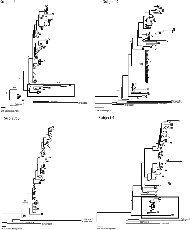
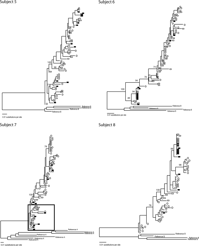
Understanding the dynamics and spread of human immunodeficiency virus type 1 (HIV-1) within the body, including within the female genital tract with its central role in heterosexual and peripartum transmission, has important implications for treatment and vaccine development. To study HIV-1 populations within tissues, we compared viruses from across the cervix to those in peripheral blood mononuclear cells (PBMC) during effective and failing antiretroviral therapy (ART) and in patients not receiving ART. Single-genome sequences of the C2-V5 region of HIV-1 env were derived from PBMC and three cervical biopsies per subject. Maximum-likelihood phylogenies were evaluated for differences in genetic diversity and compartmentalization within and between cervical biopsies and PBMC. All subjects had one or more clades with genetically identical HIV-1 env sequences derived from single-genome sequencing. These sequences were from noncontiguous cervical biopsies or from the cervix and circulating PBMC in seven of eight subjects. Compartmentalization of virus between genital tract and blood was observed by statistical methods and tree topologies in six of eight subjects, and potential genital lineages were observed in two of eight subjects. The detection of monotypic sequences across the cervix and blood, especially during effective ART, suggests that cells with provirus undergo clonal expansion. Compartmentalization of viruses within the cervix appears in part due to viruses homing to and/or expanding within the cervix and is rarely due to unique viruses evolving within the genital tract. Further studies are warranted to investigate mechanisms producing monotypic viruses across tissues and, importantly, to determine whether the proliferation of cells with provirus sustain HIV-1 persistence in spite of effective ART.
Figures
References
-
- Bailey, J. R., A. R. Sedaghat, T. Kieffer, T. Brennan, P. K. Lee, M. Wind-Rotolo, C. M. Haggerty, A. R. Kamireddi, Y. Liu, J. Lee, D. Persaud, J. E. Gallant, J. Cofrancesco, Jr., T. C. Quinn, C. O. Wilke, S. C. Ray, J. D. Siliciano, R. E. Nettles, and R. F. Siliciano. 2006. Residual human immunodeficiency virus type 1 viremia in some patients on antiretroviral therapy is dominated by a small number of invariant clones rarely found in circulating CD4+ T cells. J. Virol. 806441-6457. - PMC - PubMed
-
- Beerli, P., N. C. Grassly, M. K. Kuhner, D. Nickle, O. Pybus, M. Rain, A. Rambaut, A. G. Rodrigo, and Y. Wang. 2001. Population genetics of HIV: parameter estimation using genealogy-based methods, p. 217-258. In A. G. Rodrigo and G. H. Learn (ed.), Computational and evolutionary analyses of HIV sequences. Kluwer Academic Publishers, Boston, MA.
-
- Chang, J. T., V. R. Palanivel, I. Kinjyo, F. Schambach, A. M. Intlekofer, A. Banerjee, S. A. Longworth, K. E. Vinup, P. Mrass, J. Oliaro, N. Killeen, J. S. Orange, S. M. Russell, W. Weninger, and S. L. Reiner. 2007. Asymmetric T lymphocyte division in the initiation of adaptive immune responses. Science 3151687-1691. - PubMed
-
- Cheynier, R., S. Henrichwark, F. Hadida, E. Pelletier, E. Oksenhendler, B. Autran, and S. Wain-Hobson. 1994. HIV and T cell expansion in splenic white pulps is accompanied by infiltration of HIV-specific cytotoxic T lymphocytes. Cell 78373-387. - PubMed
Publication types
MeSH terms
Substances
Grants and funding
LinkOut - more resources
Full Text Sources
Miscellaneous
