. 2009 Jun;83(12):6269-78.
doi: 10.1128/JVI.00318-09. Epub 2009 Apr 1.

Adeno-associated virus replication induces a DNA damage response coordinated by DNA-dependent protein kinase

Affiliations

Adeno-associated virus replication induces a DNA damage response coordinated by DNA-dependent protein kinase

Rachel A Schwartz et al. J Virol. 2009 Jun.

Abstract

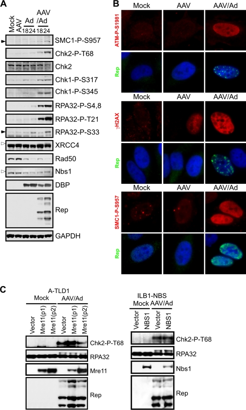
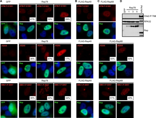
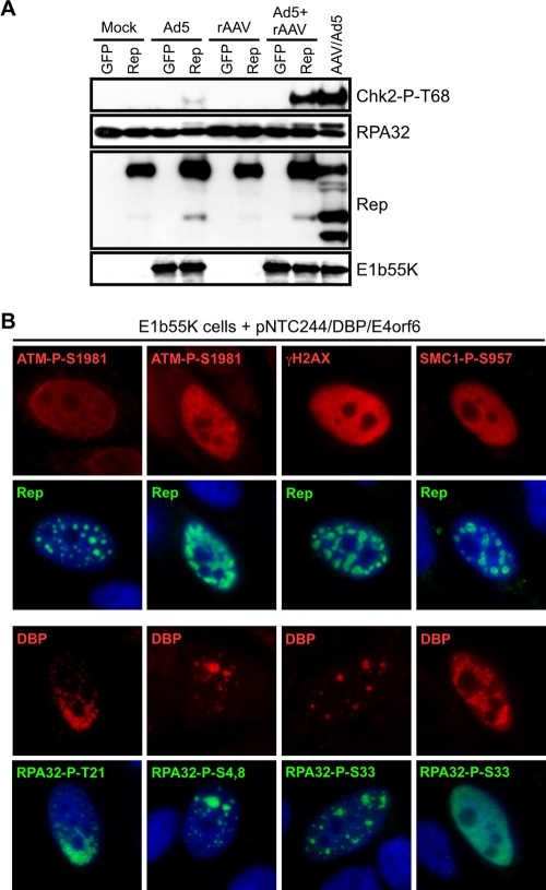
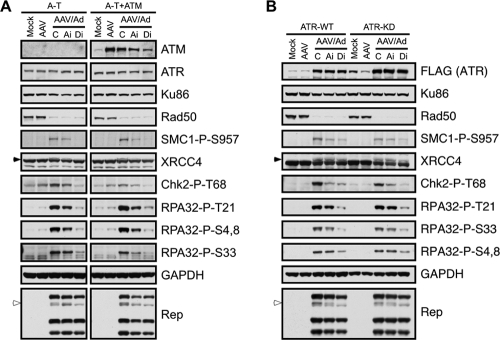
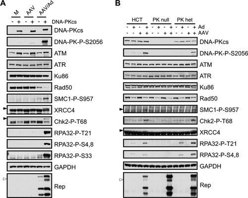
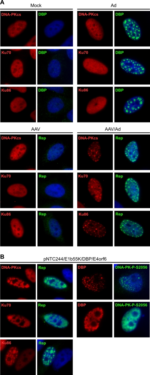
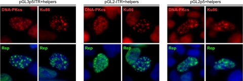
The parvovirus adeno-associated virus (AAV) contains a small single-stranded DNA genome with inverted terminal repeats that form hairpin structures. In order to propagate, AAV relies on the cellular replication machinery together with functions supplied by coinfecting helper viruses such as adenovirus (Ad). Here, we examined the host cell response to AAV replication in the context of Ad or Ad helper proteins. We show that AAV and Ad coinfection activates a DNA damage response (DDR) that is distinct from that seen during Ad or AAV infection alone. The DDR was also triggered when AAV replicated in the presence of minimal Ad helper proteins. We detected autophosphorylation of the kinases ataxia telangiectasia mutated (ATM) and DNA-dependent protein kinase catalytic subunit (DNA-PKcs) and signaling to downstream targets SMC1, Chk1, Chk2, H2AX, and XRCC4 and multiple sites on RPA32. The Mre11 complex was not required for activation of the DDR to AAV infection. Additionally, we found that DNA-PKcs was the primary mediator of damage signaling in response to AAV replication. Immunofluorescence revealed that some activated damage proteins were found in a pan-nuclear pattern (phosphorylated ATM, SMC1, and H2AX), while others such as DNA-PK components (DNA-PKcs, Ku70, and Ku86) and RPA32 accumulated at AAV replication centers. Although expression of the large viral Rep proteins contributed to some damage signaling, we observed that the full response required replication of the AAV genome. Our results demonstrate that AAV replication in the presence of Ad helper functions elicits a unique damage response controlled by DNA-PK.


Figures

FIG. 1.
FIG. 1.
AAV and Ad coinfection induces an MRN-independent DDR. (A) DNA damage signaling induced by AAV and Ad coinfection. U2OS cells were infected with Ad (MOI of 25) and AAV (MOI of 2,000) alone or in combination. Cells were harvested at the indicated times and processed for immunoblotting. Open arrowheads indicate slower-migrating phosphorylated proteins and closed arrowheads indicate the phosphorylated protein of interest. GAPDH served as a loading control. (B) HeLa cells were infected with the indicated viruses for approximately 20 h before being fixed and processed for immunofluorescence. Cells were stained with the indicated antibodies to mark activated DDR proteins and Rep centers, and DAPI (4′,6′-diamidino-2-phenylindole) was used to stain the DNA of cell nuclei. (C) DNA damage signaling in response to AAV and Ad coinfection still occurs in cell lines lacking a functional MRN complex. A-TLD1 and NBS cells and their complemented counterparts were uninfected (Mock) or infected with AAV (MOI of 1,000) and Ad5 (MOI of 50) for 30 h. Cells were harvested and processed for immunoblotting with the indicated antibodies. Mre11-P1 and P2 represent two different pools of A-TLD1 cells transduced to express wild-type Mre11.
FIG. 2.
FIG. 2.
The effects of Rep proteins on DNA damage signaling. HeLa cells were transfected with plasmids encoding GFP or Rep proteins. Cells were fixed for immunofluorescence approximately 36 h posttransfection and stained with the indicated antibodies to mark activated DDR proteins or Rep and with DAPI (4′,6′-diamidino-2-phenylindole) to stain the nuclei. At least 100 cells were quantitated for the indicated percentages of each phenotype. (A) Rep78 activates DNA damage signaling. (B) FLAG-Rep40 does not activate damage signaling. (C) Damage signaling during FLAG-Rep68 expression. (D) Rep78 does not induce downstream signaling. HeLa cells were transfected with a plasmid encoding GFP or increasing amounts of a plasmid encoding Rep78. Total DNA transfected was the same between samples and was made up with pRK5. Cells were harvested 48 h posttransfection and processed for immunoblotting with the indicated antibodies. Coinfection with AAV and Ad served as a positive control.
FIG. 3.
FIG. 3.
AAV replication induces a DNA damage response. (A) HeLa cells were either uninfected (Mock) or infected with Ad5 (MOI of 50), rAAV-GFP (MOI of 1,000), or a combination of both viruses. At 6 h postinfection, cells were transfected with plasmids encoding GFP or Rep78 and harvested 30 h posttransfection. Lysates were processed for immunoblotting against the indicated proteins. Coinfection with wild-type AAV and Ad5 served as a positive control. (B) HeLa cells transduced to express E1b55K were transfected with pNTC244 and plasmids expressing DBP and E4orf6. At approximately 36 h posttransfection, cells were fixed for immunofluorescence and stained with the indicated antibodies. AAV replication centers are marked by DBP or Rep. DAPI (4′,6′-diamidino-2-phenylindole) was used to mark cell nuclei.
FIG. 4.
FIG. 4.
ATM and ATR are not the kinases predominantly responsible for signaling in response to AAV and Ad coinfection. Immunoblotting was used to analyze AAV-induced damage signaling. Cells with individual PIKKs inactivated were either uninfected (Mock) or infected with AAV (MOI of 2,000) or AAV and Ad (MOI of 25). Infections were performed in the presence of DMSO as a control (C) or inhibitors to ATM (Ai) and DNA-PKcs (Di). Mock- and AAV-infected cells were also treated with DMSO. Cells were harvested 24 h postinfection and processed for immunoblotting with the indicated antibodies. (A) Infections in A-T cells or a matched line complemented with ATM. Right- and left-hand panels were from the same gels, and lanes not relevant to these results were removed from the figure. (B) Infections in cells expressing inducible ATR that is wild-type (ATR-WT) or kinase-dead (ATR-KD). Cells were induced to express ATR by doxycycline treatment for 48 h before infections, which then proceeded for a further 24 h. The inducible ATR protein is tagged with a FLAG epitope. GAPDH served as a loading control. The open arrowheads indicate a slower-migrating band for Rep68, and the filled arrowheads highlight specific phosphorylated proteins.
FIG. 5.
FIG. 5.
DNA-PKcs is required for signaling in response to AAV and Ad coinfection. Immunoblotting was used to analyze damage signaling to AAV and Ad coinfection in cells that lack DNA-PKcs. Cells were either uninfected or infected with AAV (MOI of 1,000 to 2,000), Ad (MOI of 25), or both viruses and harvested 24 h postinfection. Lysates were processed for immunoblotting with the indicated antibodies. The open arrowheads indicate a slower-migrating band for Rep68, and the filled arrowheads highlight specific phosphorylated proteins. (A) Infections in M059J-derived cells lines that lack DNA-PKcs (Fus9) or express the kinase (Fus1). (B) Infections in HCT-derived cells that are heterozygous or null for DNA-PKcs.
FIG. 6.
FIG. 6.
DNA-PK components localize to AAV replication centers. (A) DNA-PK components localize to viral replication centers during AAV and Ad coinfection. HeLa cells were uninfected (Mock) or infected with Ad5 (MOI of 25), AAV (MOI of 1,000), or both viruses for 24 h before fixing for immunofluorescence. Cells were stained with the indicated antibodies. Viral replication centers are detected with antibodies to DBP and Rep. DAPI (4′,6′-diamidino-2-phenylindole) marks the cell nuclei. (B) HeLa cells expressing E1b55K were transfected with pNTC244, DBP, and E4orf6 expression plasmids for approximately 36 h before cells were fixed for immunofluorescence. Viral Rep centers and DAPI staining are as described in panel A.
FIG. 7.
FIG. 7.
DNA-PK is recruited to Rep-mediated replication compartments. 293 cells were transfected with pGL-based plasmids containing the 5′ ITR (pGL2-ITR), the p5 promoter (pGL3-p5), or both elements (pGL3-p5ITR) in the presence and absence (not shown) of E4orf6, DBP, and Rep78 expression plasmids. Cells were fixed approximately 36 h posttransfection and processed for immunofluorescence with the indicated antibodies. Rep marks replication centers induced by all pGL-based plasmids, and DAPI (4′,6′-diamidino-2-phenylindole) stains the cell nuclei.

References

    1. Alexander, I. E., D. W. Russell, and A. D. Miller. 1994. DNA-damaging agents greatly increase the transduction of nondividing cells by adeno-associated virus vectors. J. Virol. 688282-8287. - PMC - PubMed
    1. Bakkenist, C. J., and M. B. Kastan. 2003. DNA damage activates ATM through intermolecular autophosphorylation and dimer dissociation. Nature 421499-506. - PubMed
    1. Berthet, C., K. Raj, P. Saudan, and P. Beard. 2005. How adeno-associated virus Rep78 protein arrests cells completely in S phase. Proc. Natl. Acad. Sci. USA 10213634-13639. - PMC - PubMed
    1. Boyer, J., K. Rohleder, and G. Ketner. 1999. Adenovirus E4 34k and E4 11k inhibit double strand break repair and are physically associated with the cellular DNA-dependent protein kinase. Virology 263307-312. - PubMed
    1. Branzei, D., and M. Foiani. 2008. Regulation of DNA repair throughout the cell cycle. Nat. Rev. Mol. Cell Biol. 9297-308. - PubMed

Publication types