. 2009 Jun;83(12):6171-83.
doi: 10.1128/JVI.02163-08. Epub 2009 Apr 1.

The virological synapse facilitates herpes simplex virus entry into T cells

Affiliations

The virological synapse facilitates herpes simplex virus entry into T cells

Martine Aubert et al. J Virol. 2009 Jun.

Abstract

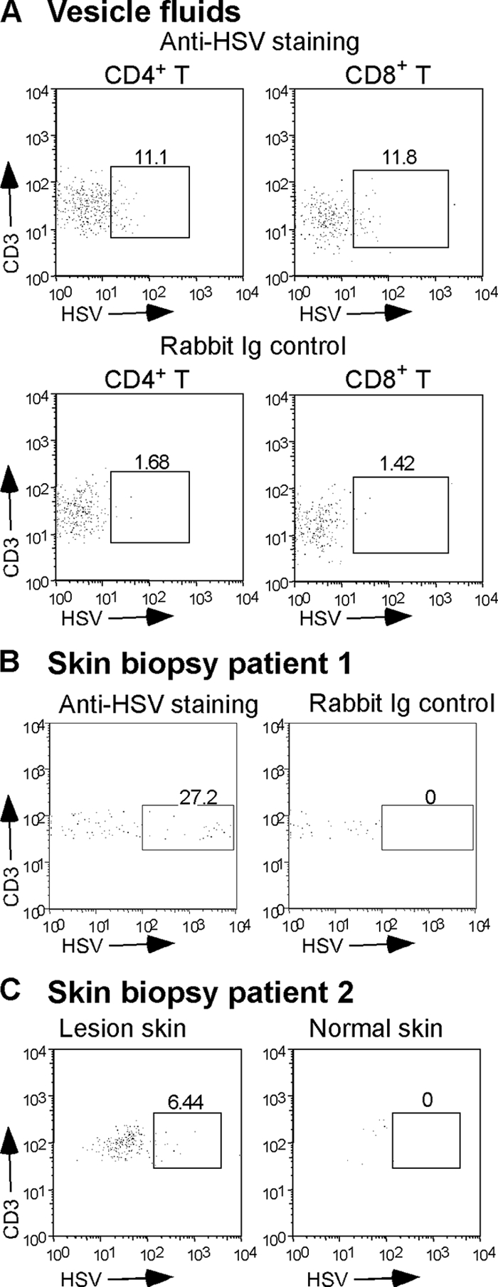
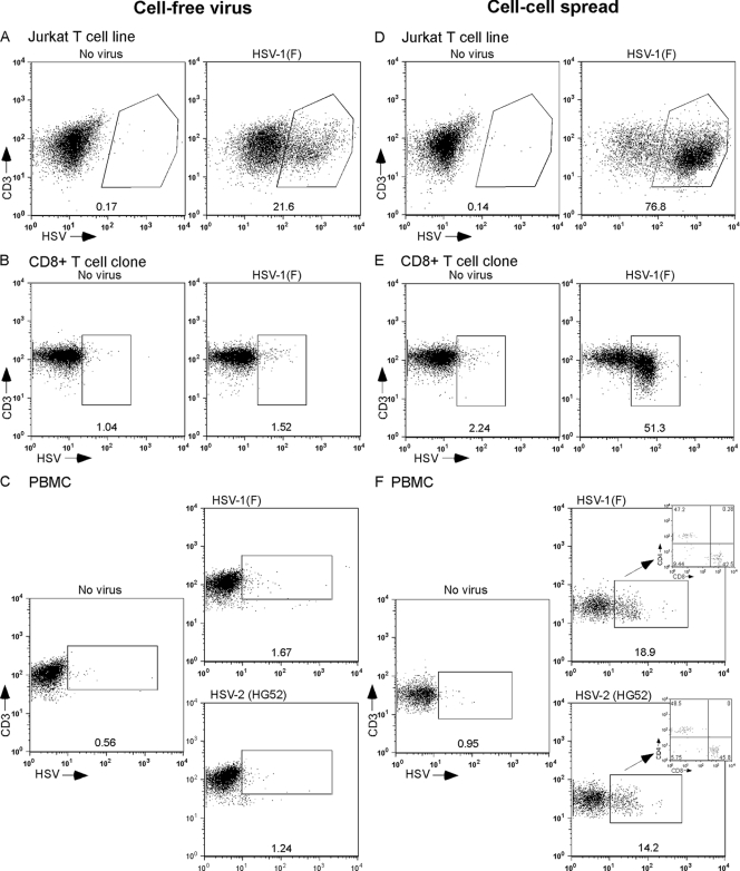
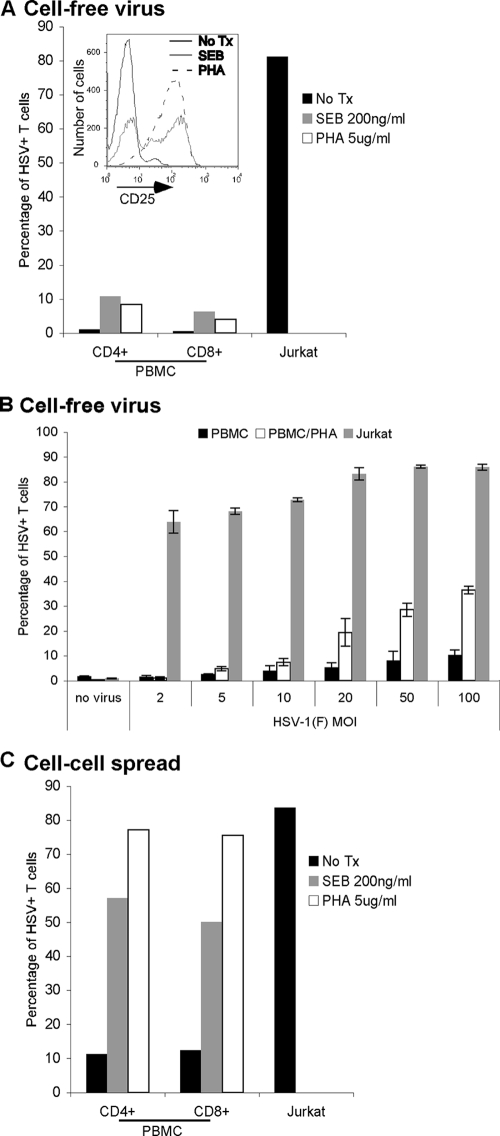
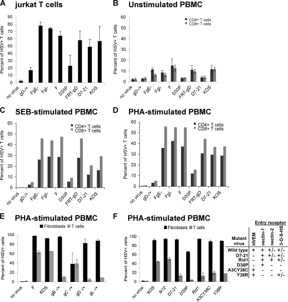
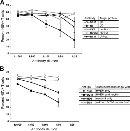
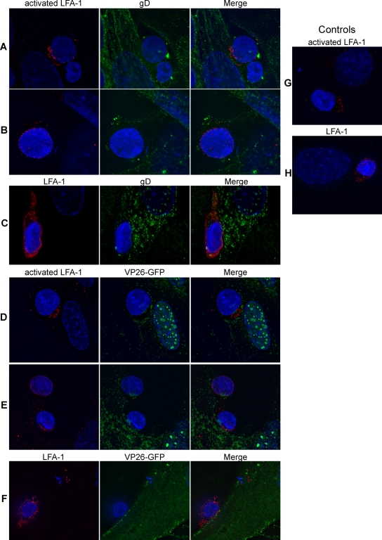
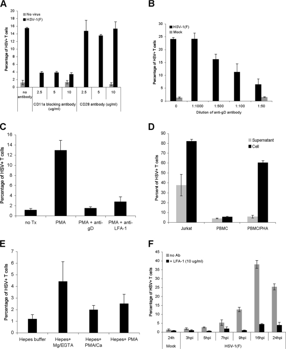
The virological synapse (VS) is a specialized molecular structure that facilitates the transfer of certain lymphotropic viruses into uninfected T cells. However, the role of the VS in the transfer of nonlymphotropic viruses into T cells is unknown. Herpes simplex virus (HSV) has been shown in vitro to infect T cells and modulate T-cell receptor function, thereby suppressing T-cell antiviral function. However, whether such infection of T cells occurs in vivo is unknown. Here, we examined whether T-cell infection could be observed in human HSV disease and investigated the mechanism of HSV entry into T cells. We found that HSV-infected T cells were readily detectable during human disease, suggesting that infection and modulation of T-cell function plays a role in human immunopathology. HSV infection of both CD4(+) and CD8(+) T cells occurred much more efficiently via direct cell-to-cell spread from infected fibroblasts than by cell-free virus. Activation of T cells increased their permissivity to HSV infection. Cell-to-cell spread to T cells did not require HSV glycoproteins E and I (gE and gI), which are critical for cell-to-cell spread between epithelial cells. Transfer of HSV to T cells required gD, and the four known entry receptors appear to be contributing to viral entry, with a dominant role for the herpesvirus entry mediator and nectin-1. VS-like structures enriched in activated lymphocyte function-associated antigen 1 (LFA-1) were observed at the point of contact between HSV-infected fibroblasts and T cells. Consistent with spread occurring via the VS, transfer of HSV was increased by activation of LFA-1, and cell-to-cell spread could be inhibited by antibodies to LFA-1 or gD. Taken together, these results constitute the first demonstration of VS-dependent cell-to-cell spread for a predominantly nonlymphotropic virus. Furthermore, they support an important role for infection and immunomodulation of T cells in clinical human disease. Targeting of the VS might allow selective immunopotentiation during infections with HSV or other nonlymphotropic viruses.


Figures

FIG. 1.
FIG. 1.
HSV is detectable in T cells from human herpes lesions. (A) Vesicle fluid collected from a herpetic lesion was stained and analyzed by flow cytometry for the presence of infected T cells using MAbs to CD3, CD4, and CD8 and rabbit polyclonal anti-HSV-2. Staining control was performed using rabbit Ig control instead of anti-HSV-2. The box in each dot plot contains the T cells positive for HSV antigens; numbers represent the percentage of T cells expressing HSV antigen. (B) A biopsy of a herpetic lesion from a second patient was stained with anti-CD3 and rabbit polyclonal anti-HSV-2 or rabbit Ig control. (C) Biopsies of a herpetic lesion (lesion skin) or control uninfected skin (normal skin) from a third patient were stained for the presence of infected T cells using anti-CD3 and rabbit polyclonal anti-HSV-2.
FIG. 2.
FIG. 2.
HSV infection of human T cells occurs preferentially via cell-cell spread. The Jurkat T cell line (A and D), a CD8+ T-cell clone (B and E), or PBMC (C and F) were either infected with cell-free HSV-1(F) at an MOI of 10 at 37°C (A to C) or coincubated at 37°C with human fibroblasts infected with HSV-1(F) or HSV-2(HG52) at an MOI of 10 for 5 h at 37°C (D to F). At 16 to 18 h postinfection or exposure, cells were collected, stained for T-cell markers and HSV antigens, and analyzed by flow cytometry. The percentage of HSV-positive T cells (CD3+ HSV positive) is indicated below each window. For PBMC infected by coincubation with HSV-infected fibroblasts, the inset shows the percentage of CD4+ (upper left quadrant) and CD8+ (lower right quadrant) cells among infected CD3+ T cells.
FIG. 3.
FIG. 3.
Activation of T cells increases the efficiency of HSV infection. (A) Jurkat T cells, PBMC, or PBMC activated for 4 days at 37°C with either SEB (200 ng/ml) or PHA (5 μg/ml) were infected with cell-free HSV-1(F) at an MOI of 10 for 16 to 18 h at 37°C, stained for T-cell markers (CD3, CD4, and CD8) and HSV antigens, and analyzed by flow cytometry. Bars indicate the percentage of T cells (CD3+) positive for HSV antigens. The inset shows flow cytometry analysis of T cells (CD3+) for CD25 prior to infection of either untreated (no Tx) PBMC or SEB- or PHA-activated PBMC. (B) Jurkat T cells, PBMC, or PBMC activated for 2 days with PHA (5 μg/ml) were infected with cell-free HSV-1(F) at the indicated MOI for 24 h at 37°C, stained for CD3 and HSV antigens, and analyzed by flow cytometry. Bars indicate the percentage of T cells (CD3+) positive for HSV antigens. (C) Jurkat T cells, PBMC, or PBMC activated for 4 days at 37°C with either SEB (200 ng/ml) or PHA (5 μg/ml) were coincubated for 16 to 18 h at 37°C with human fibroblasts infected with HSV-1(F) at an MOI of 10 for 5 h at 37°C, stained for T-cell markers (CD3, CD4, and CD8) and HSV antigens, and analyzed by flow cytometry. Bars represent the percentage of T cells (CD3+ cells) positive for HSV antigens. Panels A and C show the results from one representative experiment, and panel B shows the mean ± standard deviation of triplicate wells for each condition from one experiment.
FIG. 4.
FIG. 4.
gD, but not gE or gI, is required for cell-cell spread of HSV to T cells. Jurkat T cells (A), unstimulated PBMC (B), or PBMC activated with either SEB (C) or PHA (D, E, and F) were coincubated at 37°C for either 16 to 18 h with uninfected human fibroblasts (no virus) or fibroblasts infected at an MOI of 10 for 5 h at 37°C with the indicated viruses (A to D); alternatively, cells were coincubated for 2 h with uninfected human fibroblasts (no virus) or fibroblasts infected at an MOI of 10 for 16 h at 37°C with the indicated viruses, removed, washed with citrate buffer and PBS, and further incubated in culture medium at 37°C for 24 h (E and F). After collection, the T cells were stained either for both HSV antigens and CD3 (A, E, and F) or for HSV antigens, CD3, CD4, and CD8 (B to D), followed by flow cytometry analysis. The graphs show the percentage of CD3+ T cells positive for HSV antigens (A, E, and F) or CD3+ CD4+ (black bars) and CD3+ CD8+ (gray bars) T cells positive for HSV antigens (B to D). In panels E and F, additional wells of fibroblasts were infected in parallel with the wells of infected fibroblasts used for the cell spread experiment; fibroblasts were collected at the end of the coincubation period of the cell spread experiment and stained for HSV antigens and analyzed by flow cytometry. Black bars indicate the percent infection of fibroblasts; gray bars indicate the percent infection of T cells. The table indicates the ability (+) or inability (−) of mutant viruses to use the known HSV entry receptors. HSV(F) is the parental virus for FgE− (deletion of gE), FgI− (deletion of gI), and gD−/+; FRT-gD is the parental virus for D30P and Δ7-21; KOS is the parental virus for A3CY38C, Y38R, and the gB, gC, and gL mutants in panel E. Panels A and B show the average of two independent experiments, panel C or D shows the results from one experiment, and panels E and F show the results of one representative experiment with duplicate wells.
FIG. 5.
FIG. 5.
Blocking of HSV spread with either antibody directed against known HSV entry receptors or HSV glycoproteins. (A) PBMC stimulated with PHA (1 μg/ml for 2 days at 37°C) were coincubated for 2 h at 37°C with fibroblasts infected with HSV-1(F) at an MOI of 10 for 16 h in the presence of various dilutions of antibody. For anti-HVEM and anti-nectin-1, the diluted antibodies were added to the PBMC and incubated for 20 min at 37°C before coincubation with the infected fibroblasts. For anti-gB, anti-gD, and anti-gH-gL, the diluted antibodies were added to the infected fibroblasts and incubated for 20 min at 37°C before coincubation with the PBMC. After the 2-h coincubation, the T cells were removed, washed with citrate buffer and PBS, further incubated at 37°C in culture medium for 24 h, stained for CD3 and HSV antigens, and analyzed by flow cytometry. The table indicates the reactivity of the different antibodies used in the experiment. Antibody binding and the ability to block infection were confirmed by direct infection of control HEp-2 cells in the presence of antibody and ranged from ∼90% to ∼40% inhibition of infection relative to cells in the absence of antibody (R8 > SS10 > R137 ≈ CK41 ≈ HVEM) (data not shown). (B) PBMC stimulated with PHA as described in panel A were coincubated for 2 h at 37°C with fibroblasts infected with HSV-1(F) at an MOI of 10 for 16 h in the presence of MAb dilutions directed against different gD epitopes. The diluted antibodies were added to the infected fibroblasts and incubated for 20 min at 37°C before coincubation with the PBMC. After the 2-h coincubation, the T cells were removed, washed with citrate buffer and PBS, further incubated at 37°C in culture medium for 24 h, stained for CD3 and HSV antigens, and analyzed by flow cytometry. The table indicates the ability of the different MAbs to block gD binding to HVEM and/or nectin-1. Antibody binding and the ability to block infection were confirmed by performing direct infection of control HEp-2 cells in the presence of antibody and ranged from ∼90% to ∼20% inhibition of infection (DL11 > 1D3 ≈ HD1 ≈ DL6) (data not shown). Both panels show results from duplicate wells from one representative of two independent experiments.
FIG. 6.
FIG. 6.
Detection of LFA-1 at the site of contact between HSV-infected fibroblasts and T cells. (A to C) PBMC stimulated with PHA (5 μg/ml for 2 days at 37°C) were coincubated for 1 h at 37°C with fibroblasts infected with HSV-1(F) at an MOI of 2 for 16 h, fixed, and stained for either active LFA-1 (red) and gD (green) (A and B) or total LFA-1 (red) and gD (green) (C). (D to F) PBMC stimulated with PHA as above were coincubated for 1 h at 37°C with fibroblasts infected with HSV-1(K26GFP) at an MOI of 2 for 16 h, fixed, and stained for either active LFA-1 (red) (D and E) or total LFA-1 (red) (F). K26GFP produces and incorporates into the virion the capsid protein VP26 fused to GFP (VP26-GFP). (G and H) PBMC stimulated with PHA as above were coincubated for 1 h at 37°C with uninfected fibroblasts, fixed, and stained for either active LFA-1 (red) (G) or total LFA-1 (red) (H). In all panels, the nuclei were stained with Hoechst.
FIG. 7.
FIG. 7.
Role of the VS during HSV cell-cell spread to T cells. (A) A CD8+ T-cell clone was preincubated for 20 min at 37°C with either anti-LFA-1 blocking antibody (CD11a) or anti-CD28 antibody at the indicated concentrations before addition to uninfected fibroblasts (no virus) or fibroblasts infected with HSV-1(F) at an MOI of 10 for 16 h at 37°C. After 2 h at 37°C, the T cells were removed, washed with citrate buffer, further incubated at 37°C for 16 h, stained for CD3 and HSV antigens, and then analyzed by flow cytometry. (B) A CD8+ T-cell clone was coincubated at 37°C with mock-infected fibroblasts or fibroblasts infected with HSV-1(F) at an MOI of 10 for 16 h at 37°C in the absence or presence of neutralizing anti-gD antibody at the indicated dilutions. After 2 h, the T cells were removed, washed with citrate buffer, further incubated at 37°C for 16 h, stained for CD3 and HSV antigens, and then analyzed by flow cytometry. (C) PBMC were incubated for 20 min at 37°C with either culture medium (untreated; no Tx), culture medium with PMA (50 ng/ml), or culture medium with PMA and anti-LFA-1 blocking antibody (PMA+anti-LFA-1), and then added for 2 h to fibroblasts infected with HSV-1(F) at an MOI of 10 for 16 h. In one group the HSV-infected fibroblasts were preincubated with neutralizing anti-gD antibody (1:50 dilution) for 20 min prior to the addition of PMA-stimulated PBMC (PMA+anti-gD). After the 2-h coincubation, the T cells were removed, washed with citrate buffer and PBS, further incubated at 37°C in culture medium for 24 h, stained for CD3 and HSV antigens, and analyzed by flow cytometry. (D) Jurkat T cells, unstimulated PBMC, and PHA-activated PBMC (5 μg/ml 2 days at 37°C) were exposed for 2 h at 37°C to either fibroblasts that were infected with HSV-1(F) at an MOI of 10 for 16 h at 37°C and washed with PBS prior to the addition of T cells (Cell) or the medium (16 to 18 h postinfection) collected from fibroblasts that were infected with HSV-1(F) at an MOI of 10 for 16 h, washed with PBS, and incubated in medium for 2 h (16 to 18 h postinfection) (Supernatant). In both cases after a 2-h incubation, T cells were removed, spun, washed with citrate buffer and PBS, further incubated at 37°C for 24 h, stained for CD3 and HSV antigens, and analyzed by flow cytometry. (E) PBMC were resuspended for 30 min in either HEPES buffer, HEPES buffer plus Mg2+-EGTA, HEPES buffer plus PMA-Ca2+, or HEPES buffer plus PMA and then incubated for 1 h at 37°C with fibroblasts infected with HSV-1(F) at an MOI of 10 for 16 h. At the end of the coincubation, the PBMC were removed, washed with citrate buffer and PBS, further incubated in culture medium for 24 h, stained for CD3 and HSV antigens, and analyzed by flow cytometry. (F) PHA-activated PBMC (5 μg/ml 2 days at 37°C) were added for 2 h at 37°C to either uninfected fibroblasts (24 h; Mock) or HSV-infected fibroblasts (MOI of 10) at different times postinfection in the absence (no Ab) or presence of anti-LFA-1 blocking antibody (+LFA-1); PBMC were then removed, washed with citrate buffer and PBS, further incubated at 37°C for 20 to 25 h, stained for CD3 and HSV antigens, and then analyzed by flow cytometry. hpi, hours postinfection.

References

    1. Arthos, J., C. Cicala, E. Martinelli, K. Macleod, D. Van Ryk, D. Wei, Z. Xiao, T. D. Veenstra, T. P. Conrad, R. A. Lempicki, S. McLaughlin, M. Pascuccio, R. Gopaul, J. McNally, C. C. Cruz, N. Censoplano, E. Chung, K. N. Reitano, S. Kottilil, D. J. Goode, and A. S. Fauci. 2008. HIV-1 envelope protein binds to and signals through integrin α4β7, the gut mucosal homing receptor for peripheral T cells. Nat. Immunol. 9301-309. - PubMed
    1. Aubert, M., E. M. Krantz, and K. R. Jerome. 2006. Herpes simplex virus genes Us3, Us5, and Us12 differentially regulate cytotoxic T lymphocyte-induced cytotoxicity. Viral Immunol. 19391-408. - PubMed
    1. Bender, F. C., J. C. Whitbeck, H. Lou, G. H. Cohen, and R. J. Eisenberg. 2005. Herpes simplex virus glycoprotein B binds to cell surfaces independently of heparan sulfate and blocks virus entry. J. Virol. 7911588-11597. - PMC - PubMed
    1. Bouayyad, A., and J. Menezes. 1990. Comparative study of herpes simplex virus receptor expression on human lymphoid cells. Virology 179905-910. - PubMed
    1. Brickner, A. G., E. H. Warren, J. A. Caldwell, Y. Akatsuka, T. N. Golovina, A. L. Zarling, J. Shabanowitz, L. C. Eisenlohr, D. F. Hunt, V. H. Engelhard, and S. R. Riddell. 2001. The immunogenicity of a new human minor histocompatibility antigen results from differential antigen processing. J. Exp. Med. 193195-206. - PMC - PubMed

Publication types

Substances