. 2009 Apr 14;106(15):6226-31.
doi: 10.1073/pnas.0811045106. Epub 2009 Apr 1.

Activation of antibacterial autophagy by NADPH oxidases

Affiliations

Activation of antibacterial autophagy by NADPH oxidases

Ju Huang et al. Proc Natl Acad Sci U S A. .

Abstract

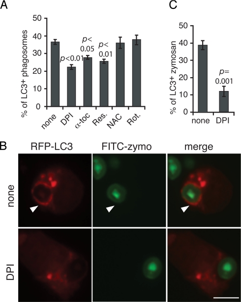
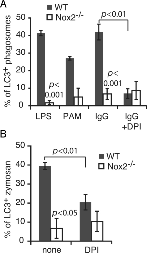
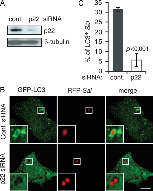
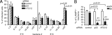
Autophagy plays an important role in immunity to microbial pathogens. The autophagy system can target bacteria in phagosomes, promoting phagosome maturation and preventing pathogen escape into the cytosol. Recently, Toll-like receptor (TLR) signaling from phagosomes was found to initiate their targeting by the autophagy system, but the mechanism by which TLR signaling activates autophagy is unclear. Here we show that autophagy targeting of phagosomes is not exclusive to those containing TLR ligands. Engagement of either TLRs or the Fcgamma receptors (FcgammaRs) during phagocytosis induced recruitment of the autophagy protein LC3 to phagosomes with similar kinetics. Both receptors are known to activate the NOX2 NADPH oxidase, which plays a central role in microbial killing by phagocytes through the generation of reactive oxygen species (ROS). We found that NOX2-generated ROS are necessary for LC3 recruitment to phagosomes. Antibacterial autophagy in human epithelial cells, which do not express NOX2, was also dependent on ROS generation. These data reveal a coupling of oxidative and nonoxidative killing activities of the NOX2 NADPH oxidase in phagocytes through autophagy. Furthermore, our results suggest a general role for members of the NOX family in regulating autophagy.


Conflict of interest statement

The authors declare no conflict of interest.

Figures

Fig. 1.
Fig. 1.
The autophagy protein LC3 is recruited to phagosomes during FcγR- and TLR-engaged phagocytosis. (A) RAW 264.7 macrophages were transiently transfected with GFP-LC3 or RFP-LC3 and then incubated with uncoated latex beads or beads coated with BSA, LPS, IgG, or FITC-zymosan (FITC-zymo) for 60 min. Arrows indicate LC3+ phagosomes and arrowheads indicate LC3+ zymosan. (Scale bar, 5 μm.) DIC, differential interference contrast. (B) The percentage of LC3+ phagosomes or zymosan (n ≥ 50) from A was quantified and compared with the group using uncoated beads. (C) Quantification of LC3+ intracellular IgG-coated beads or zymosan over time (n ≥ 80). (D) Phagosomes were isolated from RAW macrophages and fed with IgG-coated beads with or without dephenyleneiodonium chloride (DPI). Phagosome fractions were analyzed by Western blotting; 11 μg of protein from total cell lysate and 5 μg of protein from each phagosome isolate were loaded.
Fig. 2.
Fig. 2.
ROS mediate LC3 recruitment to phagosomes. RAW macrophages were transiently transfected with GFP-LC3 or RFP-LC3. Cells were incubated with IgG-coated beads or FITC-zymosan for 60 min in the absence or presence of DPI, α-tocopherol (α-toc), resveratrol (Res.), N-acetylcysteine (NAC), or rotenone (Rot.). The percentage of GFP-LC3+ intracellular beads (A) or RFP-LC3+ zymosan (C) was quantified and compared with nontreated condition. (B) Arrowhead indicates an RFP-LC3+ zymosan phagosome. (Scale bar, 5 μm.)
Fig. 3.
Fig. 3.
Autophagy targets the phagosome in a NOX2-dependent manner. (A) Bone marrow-derived neutrophils from wild-type and Nox2−/− mice were transiently transfected with GFP-LC3 and fed with latex beads coated with LPS, PAM3CSK4 (PAM), or IgG for 60 min with or without DPI treatment. The percentage of LC3+ phagosomes was quantified. Mean ± ranges (n = 2) are shown for PAM-coated beads. The mean ± SEM (n = 3) are shown for all other conditions. (B) Cells were treated the same way as in A, except they were fed with zymosan. The percentage of LC3+ zymosan was quantified and compared between wild-type and Nox2−/− neutrophils, or between ± DPI conditions.
Fig. 4.
Fig. 4.
NOX activity is required for autophagy of S. Typhimurium in epithelial cells. (A) Efficient knockdown of p22 expression in Henle-407 cells was evidenced by Western blotting. (B) GFP-LC3-transfected Henle-407 cells were treated with control or p22 siRNA. (Scale bar, 10 μm.) Insets are magnified images of selected regions. (C) The percentage of LC3+ intracellular bacteria from B was quantified.
Fig. 5.
Fig. 5.
NOX-dependent autophagy restricts S. Typhimurium replication in epithelial cells. (A) Henle-407 cells were treated with control, p22, or ATG12 siRNA for 48 hr, then infected with RFP-Sal for 2 and 8 hr. Bacterial number per infected cell was quantified and categorized into 5 categories (–, –, –, –, and >20). Percentage of infected cells for each category was quantified and compared with control siRNA condition. (B) Henle-407 cells were treated in the same way as described for A, except that LAMP1 was further immunostained in green. Percentage of LAMP1+ bacteria in infected cells was quantified and compared between 2 and 8 hr.

References

    1. Levine B, Deretic V. Unveiling the roles of autophagy in innate and adaptive immunity. Nat Rev Immunol. 2007;7:767–777. - PMC - PubMed
    1. Orvedahl A, Levine B. Eating the enemy within: Autophagy in infectious diseases. Cell Death Differ. 2009;16:57–69. - PMC - PubMed
    1. Park JB. Phagocytosis induces superoxide formation and apoptosis in macrophages. Exp Mol Med. 2003;35:325–335. - PubMed
    1. Sanjuan MA, et al. Toll-like receptor signalling in macrophages links the autophagy pathway to phagocytosis. Nature. 2007;450:1253–1257. - PubMed
    1. Nakatogawa H, Ichimura Y, Ohsumi Y. Atg8, a ubiquitin-like protein required for autophagosome formation, mediates membrane tethering and hemifusion. Cell. 2007;130:165–178. - PubMed

Publication types