. 2009 Jun;296(6):C1346-55.
doi: 10.1152/ajpcell.00523.2008. Epub 2009 Apr 1.

FXYD1 phosphorylation in vitro and in adult rat cardiac myocytes: threonine 69 is a novel substrate for protein kinase C

Affiliations

FXYD1 phosphorylation in vitro and in adult rat cardiac myocytes: threonine 69 is a novel substrate for protein kinase C

William Fuller et al. Am J Physiol Cell Physiol. 2009 Jun.

Abstract

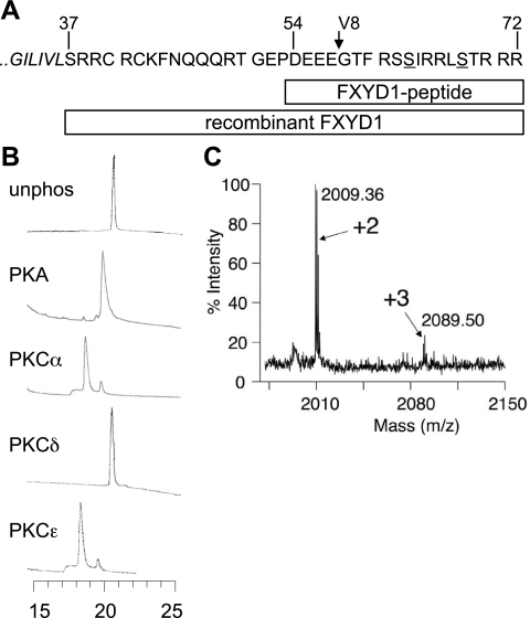
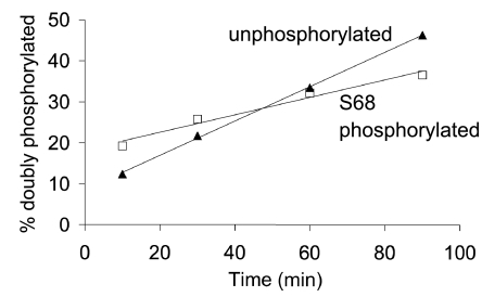
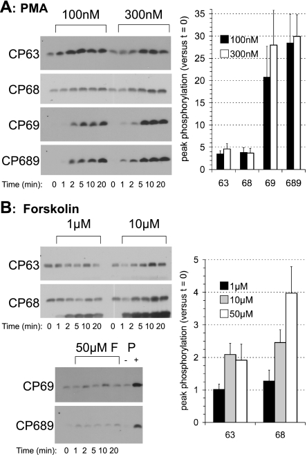
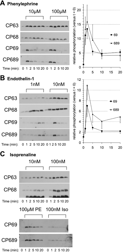
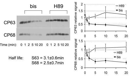
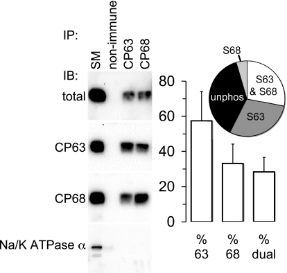
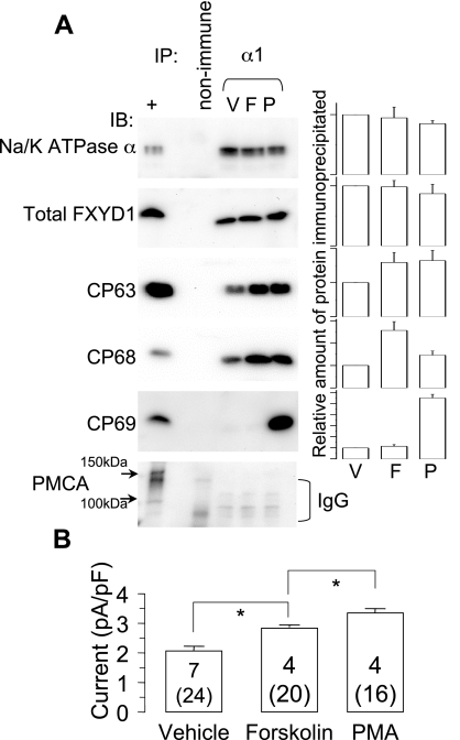
FXYD1 (phospholemman), the primary sarcolemmal kinase substrate in the heart, is a regulator of the cardiac sodium pump. We investigated phosphorylation of FXYD1 peptides by purified kinases using HPLC, mass spectrometry, and Edman sequencing, and FXYD1 phosphorylation in cultured adult rat ventricular myocytes treated with PKA and PKC agonists by phosphospecific immunoblotting. PKA phosphorylates serines 63 and 68 (S63 and S68) and PKC phosphorylates S63, S68, and a new site, threonine 69 (T69). In unstimulated myocytes, FXYD1 is approximately 30% phosphorylated at S63 and S68, but barely phosphorylated at T69. S63 and S68 are rapidly dephosphorylated following acute inhibition of PKC in unstimulated cells. Receptor-mediated PKC activation causes sustained phosphorylation of S63 and S68, but transient phosphorylation of T69. To characterize the effect of T69 phosphorylation on sodium pump function, we measured pump currents using whole cell voltage clamping of cultured adult rat ventricular myocytes with 50 mM sodium in the patch pipette. Activation of PKA or PKC increased pump currents (from 2.1 +/- 0.2 pA/pF in unstimulated cells to 2.9 +/- 0.1 pA/pF for PKA and 3.4 +/- 0.2 pA/pF for PKC). Following kinase activation, phosphorylated FXYD1 was coimmunoprecipitated with sodium pump alpha(1)-subunit. We conclude that T69 is a previously undescribed phosphorylation site in FXYD1. Acute T69 phosphorylation elicits stimulation of the sodium pump additional to that induced by S63 and S68 phosphorylation.


Figures

Fig. 1.
Fig. 1.
In vitro phosphorylation of FXYD1. A: diagrammatic representation of the FXYD1 peptides used in this study. The end of the transmembrane domain (italicized) and intracellular region of canine FXYD1 are shown, with the known phosphorylation sites S63 and S68 underlined. The site of cleavage by V8 is marked with an arrow. FXYD1 peptide was residues 54-72. The entire intracellular region of FXYD1 (37-72) was produced recombinantly. B: HPLC analysis of in vitro phosphorylated FXYD1 peptide. Peptides were phosphorylated with the kinases indicated, then analyzed on a C18 reverse-phased column. Chromatograms are temporally aligned (retention time in minutes is shown at the bottom), and absorbance at 214 nm is shown plotted against time. Phosphorylation reduces the affinity of the peptide for the C18 matrix and causes earlier elution. PKA phosphorylates the peptide at one site, PKCα and ɛ at two sites, but PKCδ not at all. C: matrix-assisted laser desorption/ionization mass spectrometry (MALDI) analysis of in vitro phosphorylated recombinant FXYD1, digested with V8. Unphosphorylated material digested with V8 yields peaks at mass-to-charge ratio (m/z) 1849.07 (58-72, containing the phosphorylation sites) and 2625.19 (37-57, not shown). Phosphorylation by PKCα shifts the peak at 1849.07 to two peaks: m/z 2009.36 (+2: theoretical m/z of dual phosphorylated peptide 2009.01) and m/z 2089.50 (+3: theoretical m/z of triply phosphorylated peptide 2088.97). Note that MALDI relies on the selection of singly charged (in this case positive) ions, meaning quantitative comparison of peak intensities of dual and triply phosphorylated species is meaningless, because less triply phosphorylated peptide will form the MH+ ion.
Fig. 2.
Fig. 2.
The effect of prephosphorylation of S68 on phosphorylation of the FXYD1 peptide by PKC. Unphosphorylated (▴) and PKA phosphorylated (□) FXYD1 peptide was phosphorylated with PKCα. Measurement of peak area of the HPLC-purified starting material ensured that substrate concentration was identical between the two reactions. Reaction progress was monitored by analyzing samples by HPLC: conversion of the starting material to a doubly phosphorylated species was measured by calculating the area of the doubly phosphorylated peak from each sample of each reaction. The percentage of peptide in each sample that had reached the doubly phosphorylated state is plotted as a function of the reaction time. The gradient of the linear regression lines are 0.21 (S68 phosphorylated starting material) and 0.42 (unphosphorylated starting material), indicating that conversion to the doubly phosphorylated state by PKCα proceeds twice as quickly for unphosphorylated starting material compared with S68 phosphorylated starting material.
Fig. 3.
Fig. 3.
Characterization of FXYD1 phosphospecific antibodies. A: the reactivity of CP63, CP68, CP69, and CP689 to in vitro phosphorylated recombinant FXYD1 and its point mutants was investigated by dot blotting in vitro phosphorylation reactions that had proceeded to completion. Antibody CP69 was preincubated with 10 μg/ml unphosphorylated blocking peptide, and antibody CP689 was preincubated with unphosphorylated blocking peptide plus two singly phosphorylated blocking peptides (all 10 μg/ml), as described in materials and methods. All phosphospecific antibodies show the appropriate reactivity, although note that mutation T69A severely limits the ability of antibody CP68 to bind to FXYD1 phosphorylated at serine 68. Antibody CP689 shows very weak binding to mutant S68A phosphorylated by PKC (i.e., phosphorylated at T69 only); however, the majority of signal generated by this antibody is for dual phosphorylated S68 and T69 [compare with signal from wild type (WT) and S63A]. B: time course of phosphorylation of wild-type recombinant FXYD1 by PKA and PKC. Phosphorylation of S63 by PKA is very slow compared with S68 (phosphorylation of S68 is complete within 10 min, but phosphorylation of S63 requires 90 min). Phosphorylation of T69 by PKCɛ severely limits binding of antibody CP68 to phosphorylated S68.
Fig. 4.
Fig. 4.
Phosphorylation of FXYD1 in cultured adult rat ventricular myocytes (ARVM) following treatment with PMA (A) and forskolin (B). PKC activation results in sustained phosphorylation of S63, S68, and T69 (A). Activation of PKA results in phosphorylation of S63 and S68, but not T69. Note that antibody CP68 cross-reacts with phospholamban phosphorylated at serine 16. A positive control [PMA-treated ARVM (P)] is shown for antibodies CP69 and CP689 in B. Representative immunoblots for 2 concentrations of agonist with each phosphospecific antibody and mean peak responses normalized to the signal at time zero (t = 0) are shown (n ≥ 6, means ± SE). F, forskolin.
Fig. 5.
Fig. 5.
Receptor-mediated FXYD1 phosphorylation in cultured ARVM. α-Adrenoceptor activation with phenylephrine (A) causes sustained phosphorylation of S63 and S68, but transient phosphorylation of T69. Endothelin type A receptor activation with endothelin-1 (B) causes qualitatively very similar effects. β-Adrenoceptor activation with isoprenaline (C) causes sustained phosphorylation of S63 and S68 but not T69. A positive control for antibodies CP69 and CP689 [phenylephrine-treated ARVM (PE)] is shown alongside isoprenaline-treated cells (Iso; C). Representative immunoblots are shown for each phosphospecific antibody. For simplicity of presentation, only mean data for antibodies CP69 and CP689 at the highest agonist dose, normalized to the signal at time zero, are shown in A and B (n ≥ 6, means ± SE).
Fig. 6.
Fig. 6.
Effect of acute inhibition of PKA and PKC on basal phosphorylation of S63 and S68 in unstimulated cultured ARVM. While the PKA inhibitor H89 (10 μM) is without effect on basal FXYD1 phosphorylation, in the presence of the PKC inhibitor bisindolylmaleimide (bis, 1 μM), FXYD1 is rapidly dephosphorylated at S63 and S68. The first 5 min of bis treatment were fitted with a single exponential, giving half-lives of 3.1 ± 0.6 min and 2.5 ± 0.7 min for phosphorylated S63 and S68, respectively.
Fig. 7.
Fig. 7.
Basal phosphorylation of S63 and S68 in cultured ARVM. Triton X-100-solubilized FXYD1 was immunoprecipitated (IP) with nonimmune serum, and antibodies CP63 and CP68, and immunoblotted (IB) with an antibody that does not distinguish between phosphorylation states (total). Immunoprecipitated FXYD1 is expressed as percentage of that found in the starting material (SM). The FXYD1/Na+-K+-ATPase α1-subunit interaction is disrupted by the presence of 1% Triton X-100 in the immunoprecipitation buffer (Na+-K+-ATPase α-subunit is not detected copurifying with FXYD1). A full explanation of the calculations of percent phosphorylation is in the text. Mean data indicating percent basal phosphorylation at each residue from 5 independent experiments are shown (means ± SE). Inset: pie chart showing the distribution of FXYD1 between phosphorylated at S63 only, phosphorylated at S68 only, dual phosphorylated (S63 and S68), and unphosphorylated states in unstimulated ARVM.
Fig. 8.
Fig. 8.
Functional effect of T69 phosphorylation on Na+-K+ pump currents. A: Na+-K+-ATPase α1-subunit was immunoprecipitated under conditions favoring copurification of FXYD1. Control immunoprecipitations used nonimmune mouse serum and untreated ARVM. +, Untreated ARVM. Representative immunoblots are shown, and the bar graphs alongside each immunoblot show the mean relative amounts of each protein immunoprecipitated, normalized to the amount purified from vehicle-treated cells (n = 5). Equal amounts of Na+-K+-ATPase α1 and copurifying total FXYD1 were immunoprecipitated from ARVM treated with vehicle [0.1% vol/vol ethanol (V)], 50 μM forskolin (F), or 300 nM PMA (P) for 10 min at 37°C. The specificity of coimmunoprecipitation was confirmed by blotting for plasma membrane Ca2+-ATPase (PMCA). We routinely observe bands at ∼140 kDa and ∼95 kDa when probing ARVM lysates for PMCA: neither form is immunoprecipitated in these reactions; however, the mouse IgG used in immunoprecipitated samples is seen. In cells treated with forskolin, copurifying FXYD1 was more phosphorylated at S63 and S68 compared with control. In cells treated with PMA, copurifying FXYD1 was more phosphorylated at S63, S68, and T69. B: cultured ARVM were treated with vehicle (0.1% vol/vol ethanol), forskolin (50 μM), PMA (300 nM). Recordings were made in the whole cell mode, with 50 mM Na+ in the pipette. Drugs were applied on the microscope stage at 35°C, and cells were left for a minimum of 5 min before establishing a seal and rupturing the membrane, to avoid disrupting signaling pathways by dialyzing the cell contents before a response developed. Recordings were made from multiple cells for up to 25 min after drugs were applied. Na+-K+ pump current was defined as the current sensitive to the removal of extracellular K. Data are means ± SE. Numbers are the number of cells (in parentheses) and number of animals (each animal gives one data point). *P < 0.05 (Student's t-test).

References

    1. Bers DM, Barry WH, Despa S. Intracellular Na+ regulation in cardiac myocytes. Cardiovasc Res 57: 897–912, 2003. - PubMed
    1. Bossuyt J, Ai X, Moorman JR, Pogwizd SM, Bers DM. Expression and phosphorylation of the Na-pump regulatory subunit phospholemman in heart failure. Circ Res 97: 558–565, 2005. - PubMed
    1. Bossuyt J, Despa S, Martin JL, Bers DM. Phospholemman phosphorylation alters its fluorescence resonance energy transfer with the Na/K-ATPase pump. J Biol Chem 281: 32765–32773, 2006. - PubMed
    1. Chibalin AV, Pedemonte CH, Katz AI, Feraille E, Berggren PO, Bertorello AM. Phosphorylation of the catalyic α-subunit constitutes a triggering signal for Na+,K+-ATPase endocytosis. J Biol Chem 273: 8814–8819, 1998. - PubMed
    1. Clerk A, Bogoyevitch MA, Anderson MB, Sugden PH. Differential activation of protein kinase C isoforms by endothelin-1 and phenylephrine and subsequent stimulation of p42 and p44 mitogen-activated protein kinases in ventricular myocytes cultured from neonatal rat hearts. J Biol Chem 269: 32848–32857, 1994. - PubMed

Publication types

MeSH terms

LinkOut - more resources