. 2009 Jun;37(10):3464-73.
doi: 10.1093/nar/gkp205. Epub 2009 Apr 1.

C19MC microRNAs are processed from introns of large Pol-II, non-protein-coding transcripts

Affiliations

C19MC microRNAs are processed from introns of large Pol-II, non-protein-coding transcripts

Marie-Line Bortolin-Cavaillé et al. Nucleic Acids Res. 2009 Jun.

Abstract

MicroRNAs are tiny RNA molecules that play important regulatory roles in a broad range of developmental, physiological or pathological processes. Despite recent progress in our understanding of miRNA processing and biological functions, little is known about the regulatory mechanisms that control their expression at the transcriptional level. C19MC is the largest human microRNA gene cluster discovered to date. This 100-kb long cluster consists of 46 tandemly repeated, primate-specific pre-miRNA genes that are flanked by Alu elements (Alus) and embedded within a approximately 400- to 700-nt long repeated unit. It has been proposed that C19MC miRNA genes are transcribed by RNA polymerase III (Pol-III) initiating from A and B boxes embedded in upstream Alu repeats. Here, we show that C19MC miRNAs are intron-encoded and processed by the DGCR8-Drosha (Microprocessor) complex from a previously unidentified, non-protein-coding Pol-II (and not Pol-III) transcript which is mainly, if not exclusively, expressed in the placenta.


Figures
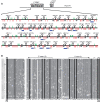
Figure 1.
Figure 1.
Revisiting microRNA gene organization at C19MC, the largest human microRNA gene cluster. (A) Schematic representation of the ∼100-kb long C19MC (HG18: 58 860 000–58 962 300) mapping at human chromosome 19q13.41. Pre-miRNA genes are symbolized as stem–loop structures. Repeated exons of C19MC-HG are indicated as grey boxes and Alus (as annotated at http://genome.ucsc.edu) are indicated by vertical bars, with green and red bars corresponding to the sense and antisense orientations relative to the pre-miRNA genes, respectively. Horizontal blue bars show miRNA gene loci that have been identified by RNA polymerase-II ChIP experiments described in Figure 3A (the number of sequenced clones is indicated in brackets). (B) Sequence alignment of human exons 1–37. The multiple sequence alignment was generated with Multalin (http://bioinfo.genopole-toulouse.prd.fr/multalin/cgi-bin/multalin.pl) and conserved nucleotides were colored with GeneDoc (http://www.Cris.com/~ketchup/genedoc.shtml). Donor (5′SS) and acceptor (3′SS) splice sites are indicated. Asterisks indicate an alternative 5′SS found in a subset of spliced exons found in cDNA clones we sequenced (not shown). The position of primers 3′-exon(1) and 5′-exon(1) used in Figure 2C are indicated on the top. An LTR sequence inserted in exon 29 has been deleted.
Figure 2.
Figure 2.
Identification of a novel, placenta-specific and repeated exon-containing noncoding RNA gene. (A) Sequence alignment of the consensus, repeated non-protein-coding exons identified in several primate species. (B) Exon–intron organization at P. anubis C19MC identified by ESTs analysis. The genomic structure of the P. anubis locus (AC184009.2 Papio BAC clone) encoding ESTs FC114510 and FC115727 is shown with sequences similar to the human C19MC exon indicated in dark grey, while similar exons not spliced in these two ESTs are indicated in light grey. The two exons exapted from inverted Alu elements are in red and pre-miRNA sequences are symbolised as stem–loop structures. Note that the 3′ exon VII (black rectangle) is similar to five ESTs at the 3′-end of the human C19MC, three of them (BF508730, BQ024418, BQ024579) are polyadenylated. For clarity, only the two exapted Alus are shown. (C) Experimental detection of (a) novel, placenta-specific mRNA-like transcript(s). Detection of spliced C19MC-HG mRNA-like transcript(s) by RT–PCR using primers as indicated in Figure 1B. (D) Schematic representation of the genomic region spanning miR-515-1 and miR-517a miRNA gene loci proposed to be transcribed by Pol-III (25). Dotted lines represent two alternative splicing events (a, b) identified by sequencing RT-PCR products using primers as indicated above exons. Other symbols are as in Figure 1. (E) Tissue-specific expression pattern of C19MC miRNAs. Total RNA from human tissues was fractionated on a denaturing 15% acrylamide/7 M urea gel and analysed by northern blot with a mixture of 32P-labelled oligonucleotides antisense to mature miR-517a, miR-515-1 and miR-519a1 that have been analysed extensively by Borchert et al. The same membrane was probed with a 5.8S rRNA probe to control gel loading. The tissue-specific expression pattern of spliced C19MC-HG transcripts was also assayed by RT–PCR using the same set of human tissues; P, primers.
Figure 3.
Figure 3.
C19MC-HG as a Pol-II, pri-miRNA transcript. (A) Pol-II is recruited at the endogenously-expressed C19MC locus. Left: miR-515-1, miR-517a and miR-519a-1 genes are endogenously-expressed in JEG3, but not in HeLa or HEK293 cells. miRNA expression was monitored by northern blot with specific oligonucleotide probes. Note that since microRNA genes mapping to C19MC are highly related to each other, causing potential cross-hybridization, it is extremely difficult to formally demonstrate their specific expression by northern blot although stringent hybridization conditions were used. Right: ChIPs were performed in JEG3 and HeLa cells with anti-Pol-II antibodies (H14, Covance) that recognize the phosphorylated CTD serine 5. Antibodies against fibrillarin (Fib) were used as a negative control. C19MC and H2B genes (used as positive control for Pol-II) were simultaneously detected by multiplexed PCR. A representative ethidium bromide-stained agarose gel of three independent ChIPs is shown and miRNA gene loci enriched for RNA Pol-II are shown in Figure 1A. (B) C19MC gene expression is sensitive to α-amanitin. JEG-3 cells were either treated by α-aminitin (left) or actinomycin D (right) as indicated and C19MC microRNA gene expression was assayed by northern blot using a mixture of 32P-labelled oligonucleotides antisense to mature miR-517a, miR-515-1 and miR-519a-1. By probing the membrane with an antisense probe to the mature miR-517 family, the same α-amanitin sensitivity was also specifically demonstrated for expression of mir-517a, one of the two C19MC microRNA genes not preceded by any (TTTT) sequence (not shown). The same membrane was also probed with a tRNATyr and pre-tRNATyr-specific probe that recognizes introns, as well as with a snaR-A specific probe. (C) C19MC-HG transcripts are processed by Microprocessor. Unspliced C19MC-HG expression was monitored by RNAse A/T1 mapping with a mixture of three related 32P-labelled antisense riboprobes to ∼180- to 200-nt long segments of C19MC spanning the pre-miRNA and the surrounding intronic sequences of the three randomly chosen miR-518b, miR-518e and miR-523 gene loci. Left: JEG-3 cells were either treated (+) or not (−) by α-aminitin (20 μg/ml, 9 h). Right: JEG-3 cells were transiently transfected with siRNA-negative control or siRNAs directed against Drosha and DGCR8 mRNAs. HeLa cells were used as a negative control. Probe: ∼2000 c.p.m. of undigested riboprobes. H20: RNAse A/T1 mapping carried out without any RNA. At the bottom of the gel, a shorter exposure is shown for pre-miRNA signals.
Figure 4.
Figure 4.
The C19MC microRNA-Alu arrangements are not consistent with robust Pol-III microRNA expression in vivo. (A) High density and strand bias of Alu repeat insertion at C19MC. Distribution of Alu repeats (histograms) and percentage of Alus in the minus strand (red curves) along C19MC. A 500 kb flanking either side of C19MC (the position of which is indicated as black arrow) was analysed. (B) Schematic representation of miRNA gene loci studied by Borchert et al. (25). Upstream Alus are indicated by boxes, with green and red boxes oriented in the sense and antisense orientations with respect to transcription of the downstream pre-miRNA genes (stem–loop structures). Alus have a dimeric structure with two related, but not equivalent, monomers: left (L) and right (R) arms. The A and B boxes of the internal Pol-III promoter are located in the left arm. Note that for miR-515-1 and miR-517a genes, the first correctly oriented Alu consists only of the right arm not expected to contain a functional Pol-III promoter. Blue and orange vertical arrows correspond to T5 (or T>5) and T4 stretches, respectively, predicted to act as Pol-III transcriptional stop signals.

References

    1. Bushati N, Cohen SM. microRNA functions. Annu. Rev. Cell Dev. Biol. 2007;23:175–205. - PubMed
    1. Kim VN. MicroRNA biogenesis: coordinated cropping and dicing. Nat. Rev. Mol. Cell Biol. 2005;6:376–385. - PubMed
    1. Kim VN, Han J, Siomi MC. Biogenesis of small RNAs in animals. Nat. Rev. Mol. Cell Biol. 2009;10:126–139. - PubMed
    1. Filipowicz W, Bhattacharyya SN, Sonenberg N. Mechanisms of post-transcriptional regulation by microRNAs: are the answers in sight? Nat. Rev. Genet. 2008;9:102–114. - PubMed
    1. Rodriguez A, Griffiths-Jones S, Ashurst JL, Bradley A. Identification of mammalian microRNA host genes and transcription units. Genome Res. 2004;14:1902–1910. - PMC - PubMed

Publication types