FGF8 signaling regulates growth of midbrain dopaminergic axons by inducing semaphorin 3F
- PMID: 19339600
- PMCID: PMC6665371
- DOI: 10.1523/JNEUROSCI.4794-08.2009
FGF8 signaling regulates growth of midbrain dopaminergic axons by inducing semaphorin 3F
Abstract
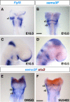
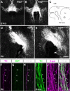
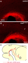
Accumulating evidence indicates that signaling centers controlling the dorsoventral (DV) polarization of the neural tube, the roof plate and the floor plate, play crucial roles in axon guidance along the DV axis. However, the role of signaling centers regulating the rostrocaudal (RC) polarization of the neural tube in axon guidance along the RC axis remains unknown. Here, we show that a signaling center located at the midbrain-hindbrain boundary (MHB) regulates the rostrally directed growth of axons from midbrain dopaminergic neurons (mDANs). We found that beads soaked with fibroblast growth factor 8 (FGF8), a signaling molecule that mediates patterning activities of the MHB, repelled mDAN axons that extended through the diencephalon. This repulsion may be mediated by semaphorin 3F (sema3F) because (1) FGF8-soaked beads induced an increase in expression of sema3F, (2) sema3F expression in the midbrain was essentially abolished by the application of an FGF receptor tyrosine kinase inhibitor, and (3) mDAN axonal growth was also inhibited by sema3F. Furthermore, mDAN axons expressed a sema3F receptor, neuropilin-2 (nrp2), and the removal of nrp-2 by gene targeting caused caudal growth of mDAN axons. These results indicate that the MHB signaling center regulates the growth polarity of mDAN axons along the RC axis by inducing sema3F.
Figures


Similar articles
-
Role of neuropilin-2 in the ipsilateral growth of midbrain dopaminergic axons.Eur J Neurosci. 2013 May;37(10):1573-83. doi: 10.1111/ejn.12190. Epub 2013 Mar 27. Eur J Neurosci. 2013. PMID: 23534961
-
Sonic hedgehog induces response of commissural axons to Semaphorin repulsion during midline crossing.Nat Neurosci. 2010 Jan;13(1):29-35. doi: 10.1038/nn.2457. Epub 2009 Nov 29. Nat Neurosci. 2010. PMID: 19946319
-
Plexin-a4 mediates axon-repulsive activities of both secreted and transmembrane semaphorins and plays roles in nerve fiber guidance.J Neurosci. 2005 Apr 6;25(14):3628-37. doi: 10.1523/JNEUROSCI.4480-04.2005. J Neurosci. 2005. PMID: 15814794 Free PMC article.
-
Fgf8 signaling for development of the midbrain and hindbrain.Dev Growth Differ. 2016 Jun;58(5):437-45. doi: 10.1111/dgd.12293. Epub 2016 Jun 7. Dev Growth Differ. 2016. PMID: 27273073 Review.
-
Induction and specification of midbrain dopaminergic cells: focus on SHH, FGF8, and TGF-beta.Cell Tissue Res. 2004 Oct;318(1):23-33. doi: 10.1007/s00441-004-0916-4. Epub 2004 Aug 19. Cell Tissue Res. 2004. PMID: 15322912 Review.
Cited by
-
Recovery from experimental parkinsonism by semaphorin-guided axonal growth of grafted dopamine neurons.Mol Ther. 2013 Aug;21(8):1579-91. doi: 10.1038/mt.2013.78. Epub 2013 Jun 4. Mol Ther. 2013. PMID: 23732989 Free PMC article.
-
RORα suppresses breast tumor invasion by inducing SEMA3F expression.Cancer Res. 2012 Apr 1;72(7):1728-39. doi: 10.1158/0008-5472.CAN-11-2762. Epub 2012 Feb 20. Cancer Res. 2012. PMID: 22350413 Free PMC article.
-
Gestational Exposure to Sodium Valproate Disrupts Fasciculation of the Mesotelencephalic Dopaminergic Tract, With a Selective Reduction of Dopaminergic Output From the Ventral Tegmental Area.Front Neuroanat. 2020 Jun 5;14:29. doi: 10.3389/fnana.2020.00029. eCollection 2020. Front Neuroanat. 2020. PMID: 32581730 Free PMC article.
-
Neural Progenitor Cells Derived from Human Embryonic Stem Cells as an Origin of Dopaminergic Neurons.Stem Cells Int. 2015;2015:647437. doi: 10.1155/2015/647437. Epub 2015 Apr 30. Stem Cells Int. 2015. PMID: 26064138 Free PMC article.
-
Crosstalk of Intercellular Signaling Pathways in the Generation of Midbrain Dopaminergic Neurons In Vivo and from Stem Cells.J Dev Biol. 2019 Jan 15;7(1):3. doi: 10.3390/jdb7010003. J Dev Biol. 2019. PMID: 30650592 Free PMC article. Review.
References
-
- Abeliovich A, Hammond R. Midbrain dopamine neuron differentiation: factors and fates. Dev Biol. 2007;304:447–454. - PubMed
-
- Ang SL. Transcriptional control of midbrain dopaminergic neuron development. Development. 2006;133:3499–3506. - PubMed
-
- Augsburger A, Schuchardt A, Hoskins S, Dodd J, Butler S. BMPs as mediators of roof plate repulsion of commissural neurons. Neuron. 1999;24:127–141. - PubMed
-
- Bally-Cuif L, Alvarado-Mallart RM, Darnell DK, Wassef M. Relationship between Wnt-1 and En-2 expression domains during early development of normal and ectopic met-mesencephalon. Development. 1992;115:999–1009. - PubMed
Publication types
MeSH terms
Substances
LinkOut - more resources
Full Text Sources
Molecular Biology Databases
Miscellaneous
