. 2009 Apr 1;29(13):4218-27.
doi: 10.1523/JNEUROSCI.4225-08.2009.

MeCP2-mediated transcription repression in the basolateral amygdala may underlie heightened anxiety in a mouse model of Rett syndrome

Affiliations

MeCP2-mediated transcription repression in the basolateral amygdala may underlie heightened anxiety in a mouse model of Rett syndrome

Megumi Adachi et al. J Neurosci. .

Abstract

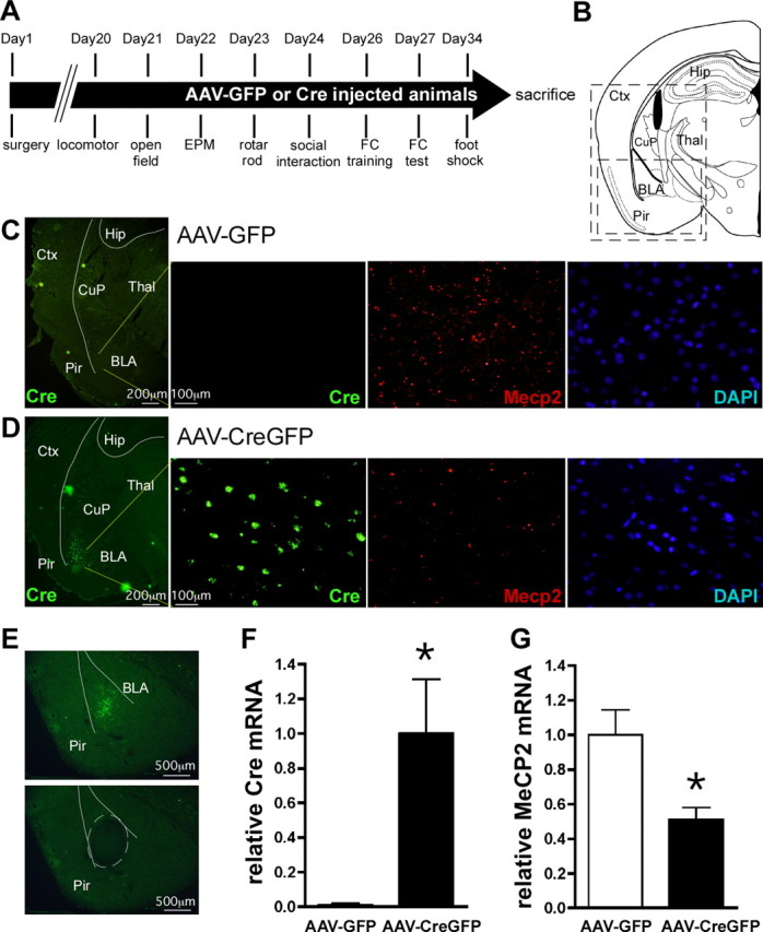
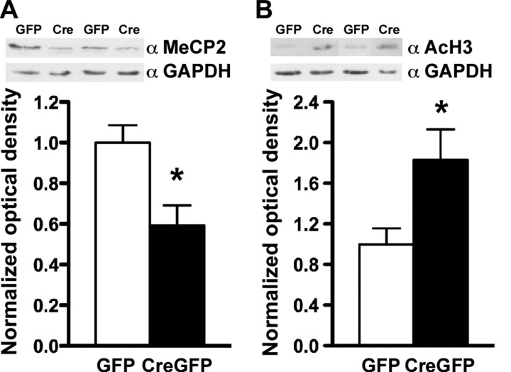
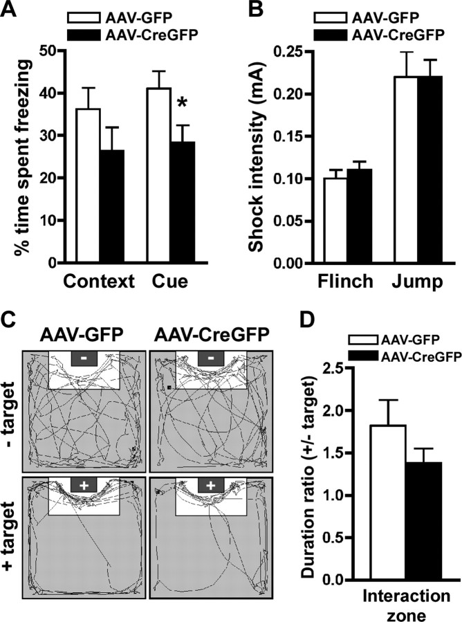
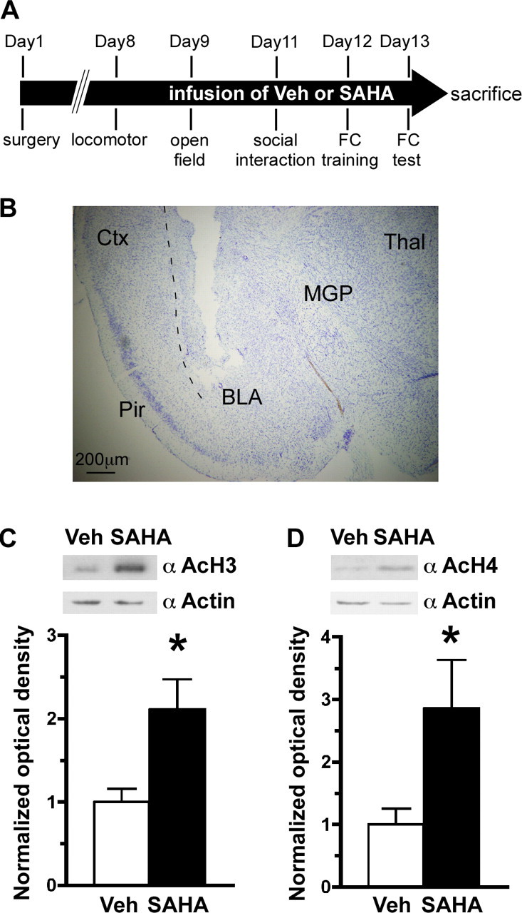
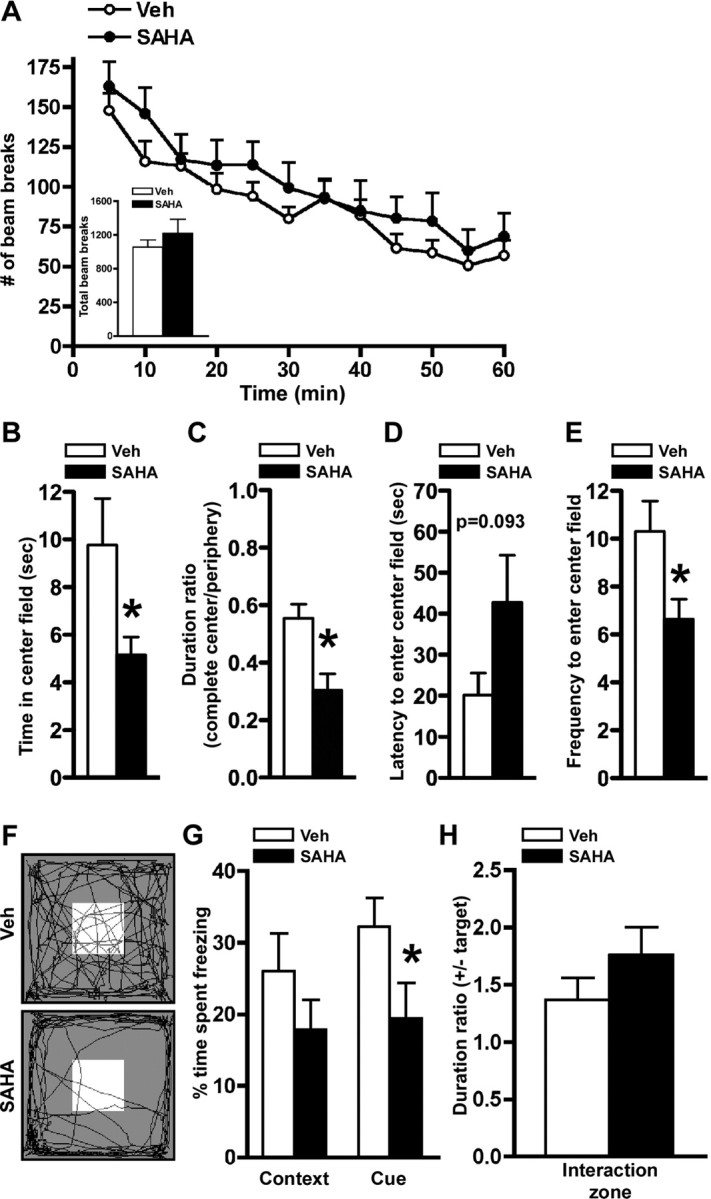
Rett syndrome (RTT) is an X-linked neurodevelopmental disorder that results from loss of function mutations in the methyl-CpG binding protein 2 (MECP2) gene. Using viral-mediated basolateral amygdala (BLA)-specific deletion of Mecp2 in mice, we show that intact Mecp2 function is required for normal anxiety behavior as well as some types of learning and memory. To examine whether these behavioral deficits are the result of impaired transcriptional repression, because Mecp2 is believed to act as a transcriptional repressor in complex with histone deacetylases (HDACs), we infused a HDAC inhibitor chronically into the BLA of wild-type mice. We found that HDAC inhibition produces behavioral deficits similar to those observed after the deletion of Mecp2 in the BLA. These results suggest a key role for Mecp2 as a transcriptional repressor in the BLA in mediating behavioral features of RTT.


Figures

Figure 1.
Figure 1.
AAV-CreGFP-mediated deletion of Mecp2 from BLA neurons. A, Experimental timeline for the behavioral experiments. B, A schematic of a mouse brain coronal section at −1.94 mm relative to bregma (adapted from Paxinos and Franklin (2001). The large box indicates the regions shown in C and D, whereas the small box represents the region shown in E. C, D, Double labeling fluorescent in situ hybridization of Cre recombinase and Mecp2 expression, visualized by FITC and Cy3 epifluorescence, respectively, in mice bilaterally injected into BLA with AAV-GFP (C) or AAV-CreGFP (D). From left, detection of Cre recombinase mRNA in the left side of a coronal section with low magnification, Cre recombinase expression, Mecp2 expression, and DAPI staining within BLA of the same field with high magnification. E, BLA containing GFP epifluorescence was laser microdissected out and subjected to real-time PCR for quantitation of Cre recombinase and Mecp2 mRNA levels. Presented is an example of the coronal section before (top) and after (bottom) laser microdissection. A microdissected region was traced with broken lines. F, G, QRT-PCR analysis of Cre recombinase (F) and Mecp2 (G) expression from laser-captured BLA. The data were normalized to 18S rRNA. *p < 0.05 by Student's t test; AAV-GFP, n = 13; AAV-CreGFP, n = 10. Ctx, Cortex; Hip, hippocampus; CuP, caudate putaman; Thal, thalamus; Pir, piriform cortex.
Figure 2.
Figure 2.
AAV-CreGFP-mediated deletion of Mecp2 from BLA neurons results in an increase in active gene transcription. Protein was extracted from the BLA region of AAV-CreGFP- and AAV-GFP-injected mice and subjected to Western blot analyses. A, AAV-CreGFP in the BLA produced a decrease (∼50%) in MeCP2 protein expression. B, Western blot analysis revealed an increase in AcH3 levels in AAV-CreGFP-injected mice compared with AAV-GFP. Optical density of MeCP2 and AcH3 was normalized to that of GAPDH. *p < 0.05 by Student's t test; AAV-GFP, n = 5; AAV-CreGFP, n = 5 for A and B.
Figure 3.
Figure 3.
BLA-specific deletion of Mecp2 results in elevated anxiety-like behavior with normal locomotion and motor coordination. A, AAV-CreGFP mice displayed motor coordination indistinguishable from that of AAV-GFP-injected mice on the rotarod test through eight trials. B, Locomotor activity was assessed by consecutive horizontal beam breaks for a 60 min testing period in 5 min increments. Inset, Total numbers of beam breaks during the test period were similar between AAV-GFP- and AAV-CreGFP-injected mice. C, D, Localized knockdown of Mecp2 in the BLA resulted in heightened anxiety-like behavior. In the elevated-plus maze test, the localized deletion of Mecp2 in the BLA resulted in a significant increase in the time spent in the closed arm (p < 0.05) (C) as well as a significant increase in the latency to enter the open arm (p < 0.05) (D), indicating an increase in anxiety-related behavior. E–H, BLA-specific Mecp2 knockdown mice were assessed for the open-field test. Presented are time in center field (E), duration ratio of time spent in complete center field to that to periphery (F), latency to enter to center field (G), and frequency to enter center field (H). Localized knockdown of Mecp2 in BLA resulted in a significant decrease in duration ratio and a significant increase in latency to enter the center field compared with AAV-GFP mice (p < 0.05), further supporting an increase in anxiety-related behavior. *p < 0.05 by Student's t test; AAV-GFP, n = 13; AAV-CreGFP, n = 10 for A–H.
Figure 4.
Figure 4.
BLA-specific deletion of Mecp2 impairs cue-dependent fear conditioning. A, The average amount of time spent freezing was determined for contextual and cue-dependent memory. AAV-CreGFP mice had a significant impairment (p < 0.05) in cue-dependent fear conditioning as demonstrated by a significant decrease in time spent freezing compared with AAV-GFP mice. There was no significant difference in context-dependent fear conditioning between AAV-GFP- and AAV-CreGFP-injected mice. B, AAV-GFP and AAV-CreGFP mice had similar footshock sensitivity to pain as measured by shock intensity to elicit flinch and jump responses. C, Representative video tracking data from the social interaction test. The gray box in the arena indicates the location of the target mouse. − and + in the gray boxes indicate the absence or presence of the target mouse, respectively. D, The ratio of time spent in the interaction zone with the target to the time without the target revealed similar levels of social interaction between the AAV-GFP and AAV-CreGFP mice. *p < 0.05 by Student's t test; AAV-GFP, n = 13; AAV-CreGFP, n = 10 for A–E.
Figure 5.
Figure 5.
Chronic infusion of an HDAC inhibitor into the BLA of C57BL/6 mice. A, Timeline of the experiment. Mice were infused for 14 d with Vehicle (Veh) or SAHA bilaterally into the BLA using osmotic pumps. B, An example of a Nissl-stained coronal brain section (left side) of the mouse through the track of a cannula. C, D, At the end of the experiment the animals were killed, placement of the cannula was confirmed, and then the BLA was dissected, and protein was isolated from the samples. Western blot analyses revealed a significant hyperacetylation of histone 3 (C) and histone 4 (D) in SAHA-infused animals. Top, Representative pictures of the Western blots. Bottom, Quantitation of the Western blots. Optical density of acetylated histone was normalized to that of actin *p < 0.05 by Student's t test; Veh, n = 10; SAHA, n = 10 for C and D. Ctx, Cortex; Thal, thalamus; MGP, medial globus pallidus; Pir, piriform cortex.
Figure 6.
Figure 6.
Mice chronically infused with SAHA in the BLA display behavioral phenotypes similar to the mice with BLA-specific deletion of Mecp2. A, Locomotor activity was unaltered by chronic SAHA infusions as assessed by consecutive horizontal beam breaks over a 60 min testing period when analyzed in 5 min increments or total beam breaks during the test period (inset). B–E, Chronic SAHA infusion resulted in increased anxiety-related behavior in the open-field test. Presented are time in center field (E), duration ratio of time spent in complete center field to that to periphery (F), latency to enter to center field (G), and frequency to enter center field (H). F, Representative video tracking data from the open-field test. A white box in the arena indicates the center field. G, In the fear-conditioning test, there was significantly less freezing in cue-dependent, but not contextual, memory formation from the mice infused with SAHA compared with those infused with vehicle (Veh). H, Chronic SAHA infusions in the BLA did not alter social interaction as assessed by the ratio of time spent in the interaction zone with the target to the time without the target. *p < 0.05 by Student's t test; Veh, n = 10; SAHA, n = 10 for A–H.

Comment in

References

    1. Adachi M, Keefer EW, Jones FS. A segment of the Mecp2 promoter is sufficient to drive expression in neurons. Hum Mol Genet. 2005;14:3709–3722. - PubMed
    1. Adachi M, Barrot M, Autry AE, Theobald D, Monteggia LM. Selective loss of brain-derived neurotrophic factor in the dentate gyrus attenuates antidepressant efficacy. Biol Psychiatry. 2008;63:642–649. - PMC - PubMed
    1. Amir RE, Van den Veyver IB, Wan M, Tran CQ, Francke U, Zoghbi HY. Rett syndrome is caused by mutations in X-linked MECP2, encoding methyl-CpG-binding protein 2. Nat Genet. 1999;23:185–188. - PubMed
    1. Asaka Y, Jugloff DG, Zhang L, Eubanks JH, Fitzsimonds RM. Hippocampal synaptic plasticity is impaired in the Mecp2-null mouse model of Rett syndrome. Neurobiol Dis. 2006;21:217–227. - PubMed
    1. Berton O, McClung CA, Dileone RJ, Krishnan V, Renthal W, Russo SJ, Graham D, Tsankova NM, Bolanos CA, Rios M, Monteggia LM, Self DW, Nestler EJ. Essential role of BDNF in the mesolimbic dopamine pathway in social defeat stress. Science. 2006;311:864–868. - PubMed

Publication types

MeSH terms