. 2009 Apr 1;29(13):4312-21.
doi: 10.1523/JNEUROSCI.0126-09.2009.

A SnoN-Ccd1 pathway promotes axonal morphogenesis in the mammalian brain

Affiliations

A SnoN-Ccd1 pathway promotes axonal morphogenesis in the mammalian brain

Yoshiho Ikeuchi et al. J Neurosci. .

Abstract

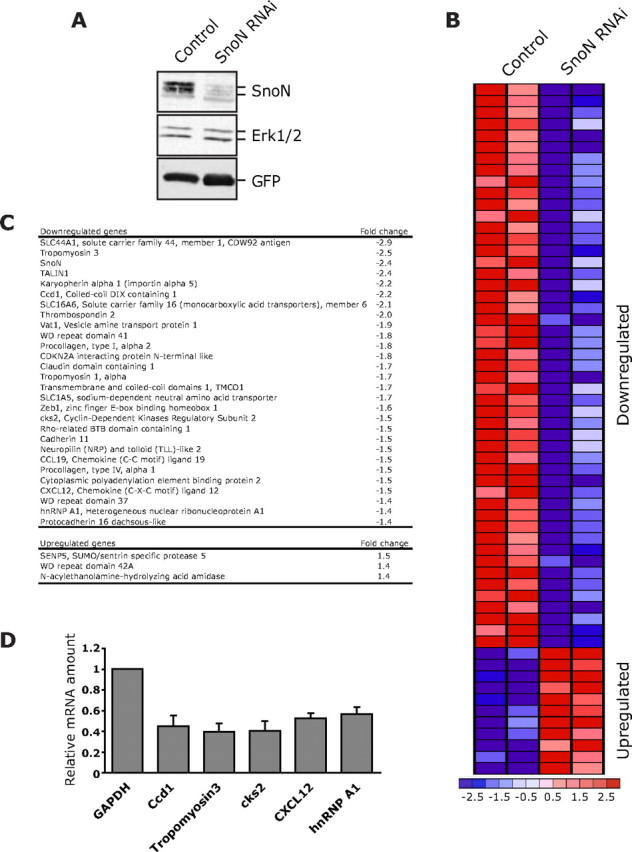
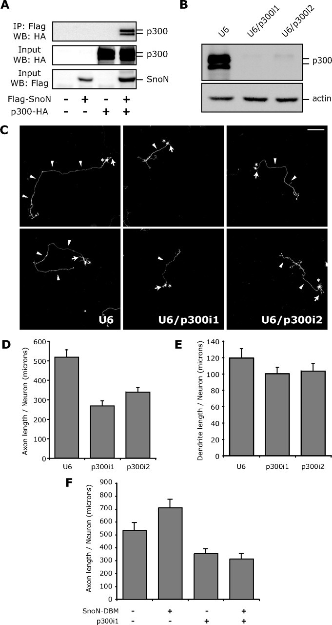
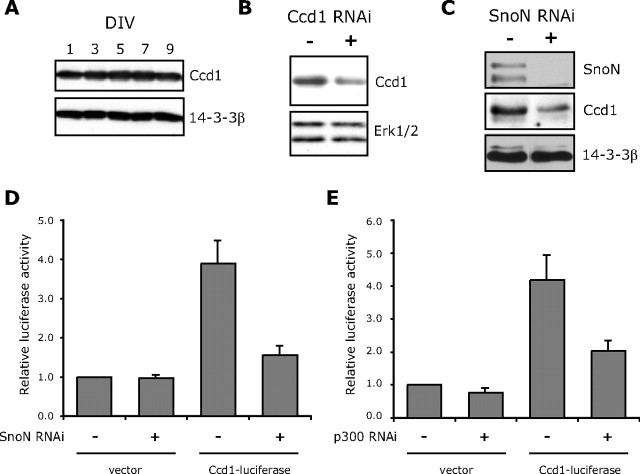
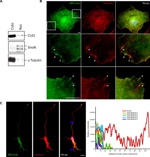
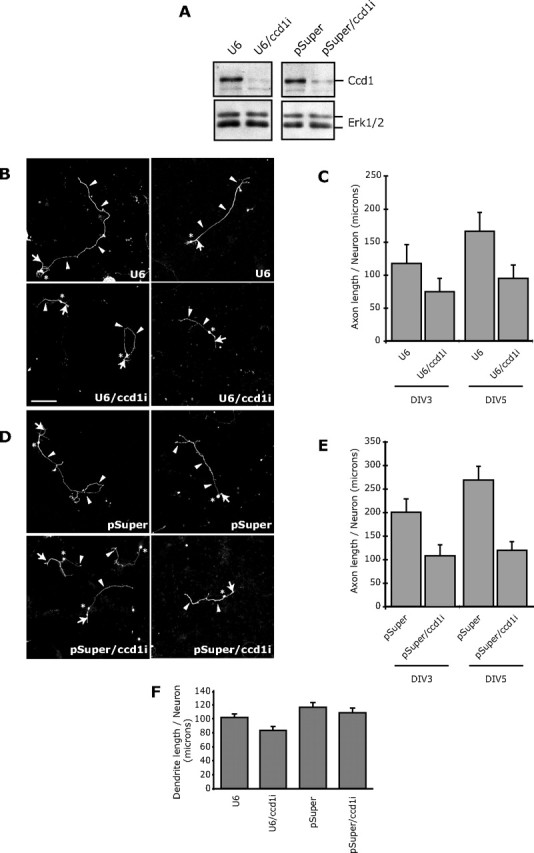
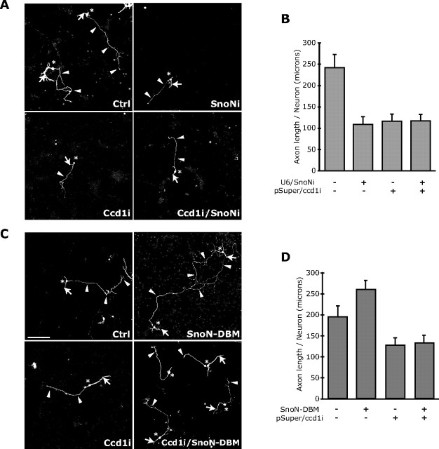
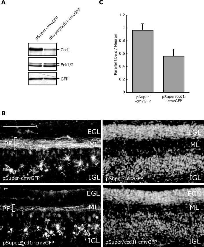
The transcriptional corepressor SnoN is a critical regulator of axonal morphogenesis, but how SnoN drives axonal growth is unknown. Here, we report that gene-profiling analyses in cerebellar granule neurons reveal that the large majority of genes altered upon SnoN knockdown are surprisingly downregulated, suggesting that SnoN may activate transcription in neurons. Accordingly, we find that the transcriptional coactivator p300 interacts with SnoN, and p300 plays a critical role in SnoN-induced axon growth. We also identify the gene encoding the signaling scaffold protein Ccd1 as a critical target of SnoN in neurons. Ccd1 localizes to the actin cytoskeleton, is enriched at axon terminals in neurons, and activates the axon growth-promoting kinase JNK (c-Jun N-terminal protein kinase). Knockdown of Ccd1 in neurons reduces axonal length and suppresses the ability of SnoN to promote axonal growth. Importantly, Ccd1 knockdown in rat pups profoundly impairs the formation of granule neuron parallel fiber axons in the rat cerebellar cortex in vivo. These findings define a novel SnoN-Ccd1 link that promotes axonal growth in the mammalian brain, with important implications for axonal development and regeneration.


Figures

Figure 1.
Figure 1.
Identification of SnoN-regulated genes in neurons. A, Lentiviral-mediated SnoN RNAi induced knockdown of endogenous SnoN in neurons. Lysates of rat cerebellar granule neurons transduced with a SnoN RNAi lentivirus that also encodes GFP or the control GFP virus were subjected to immunoblotting using the SnoN, ERK1/2, or GFP antibody. ERK1/2 served as a loading control. B, A heat map of genes altered by >1.2-fold upon SnoN knockdown compared with control virus in granule neurons. The majority (80%) of genes altered upon SnoN knockdown were downregulated. C, Selected downregulated genes (top) and upregulated genes (bottom) upon SnoN knockdown in granule neurons are listed with fold change in their expression level. D, Lysates of granule neurons transfected by a nucleofection method with the SnoN RNAi plasmid (U6/snoni) or control U6 plasmid were subjected to real-time RT-PCR to measure the mRNA levels of the indicated subset of genes. The mRNA level for each gene was normalized with glyceraldehyde-3-phosphate dehydrogenase (GAPDH) and shown as the relative ratio of normalized mRNA levels in SnoN knockdown neurons compared with control-transfected neurons. The value from three independent experiments is shown as mean + SEM. SnoN knockdown triggered the downregulation of the indicated genes.
Figure 2.
Figure 2.
The transcriptional coactivator p300 associates with SnoN and promotes axon growth. A, Lysates of 293T cells transfected with an expression plasmid encoding Flag-tagged SnoN or the control vector alone or together with an expression plasmid encoding HA-tagged p300 were immunoprecipitated (IP) using the Flag antibody followed by immunoblotting (IB) with the HA antibody. Input lysates were also immunoblotted with the Flag or HA antibody. SnoN robustly interacted with p300. B, Lysates of 293T cells transfected with the U6, U6/p300i1, or U6/p300i2 RNAi plasmid together with a plasmid expressing p300–HA were immunoblotted with the HA or actin antibody. C, Granule neurons were transfected with the control U6 (left), U6/p300i1 (middle), or U6/p300i2 RNAi plasmid (right) together with a GFP expression plasmid. Four days after transfection, granule neurons were subjected to immunocytochemistry using the GFP antibody. Neurons in which p300 knockdown was induced had shorter axons than control-transfected neurons. Arrowheads, arrows, and asterisks indicate axons, dendrites, and cell bodies, respectively. Scale bar, 100 μm. D, Morphometric analyses of granule neurons transfected and analyzed as in C. Quantification of axon length is shown as mean + SEM. Axon length was significantly reduced in p300 knockdown neurons compared with control-transfected neurons (ANOVA, p < 0.05). Total number of neurons measured = 267. E, Granule neurons transfected with the U6, U6/p300i1, or U6/p300i2 RNAi plasmid together with the GFP expression plasmid were subjected to immunocytochemistry using the GFP antibody followed by morphometric analysis of total dendrite length. No significant difference in total dendrite length between p300 knockdown and control-transfected neurons was found. Total number of neurons measured = 149. F, Granule neurons transfected with the control U6 or U6/p300i1 RNAi plasmid together with the SnoN–DBM expression plasmid or control vector were analyzed as in D. SnoN–DBM expression increased axon length compared with control-transfected neurons (ANOVA; p < 0.05). Knockdown of p300 significantly reduced axon length in the background of SnoN–DBM expression (ANOVA; p < 0.05). Total number of neurons measured = 218 neurons. These results show that p300 knockdown overrides the ability of SnoN–DBM to stimulate axon growth.
Figure 3.
Figure 3.
Identification of Ccd1 as a SnoN-regulated downstream target gene. A, Lysates of granule neurons cultured for the indicated DIV were immunoblotted with the Ccd1 or 14-3-3β antibody. Cultured days in vitro are indicated. B, Lysates of granule neurons transfected by nucleofection method with the Ccd1 RNAi plasmid (pSuper/ccd1i) or control pSuper plasmid were immunoblotted with the Ccd1 or ERK1/2 antibody. Ccd1 RNAi reduced the Ccd1-immunoreactive band, specifically. C, Lysates of granule neurons transfected by nucleofection with the SnoN RNAi (U6/snoni) or control U6 plasmid were immunoblotted with the SnoN, Ccd1, or 14-3-3β antibody. SnoN knockdown triggered downregulation of Ccd1 protein in neurons. D, Granule neurons were transfected with the SnoN RNAi (U6/snoni) or control U6 plasmid together with the Ccd1–luciferase reporter or the control vector pGL3basic and the EF renilla reporter, the latter serving as an internal control for transfection efficiency. The level of Ccd1–luciferase reporter gene expression was significantly reduced in SnoN knockdown neurons compared with control-transfected neurons (ANOVA; p < 0.01; n = 3). E, Granule neurons were transfected with the p300 RNAi (U6/p300i1) or control U6 plasmid together with the Ccd1–luciferase reporter or the control vector pGL3basic and the EF renilla reporter, the latter serving as an internal control for transfection efficiency. The level of Ccd1–luciferase reporter gene expression was significantly reduced in p300 knockdown neurons compared with control-transfected neurons (ANOVA; p < 0.05; n = 3).
Figure 4.
Figure 4.
Ccd1 is localized in the cytoplasm and enriched at axon terminals in neurons. A, Lysates of granule neurons were fractionated into cytoplasmic (Cyto) and nuclear (Nuc) fractions and then immunoblotted with the Ccd1, SnoN, or α-tubulin antibody. SnoN and α-tubulin served as markers of the nuclear and the cytoplasmic fractions, respectively. B, COS cells were transfected with the GFP–Ccd1 expression plasmid and subjected to immunocytochemistry using the GFP antibody (left), rhodamine–phalloidin staining (middle), or the DNA dye bisbenzimide (Hoechst, merged with GFP and phalloidin staining in right panels). Boxed regions in the top left panel are enlarged in second and third row. GFP–Ccd1 colocalizes with phalloidin-positive fibers, suggesting that Ccd1 is localized at the actin cytoskeleton as indicated with arrowheads. Scale bar, 10 μm. C, Hippocampal neurons transfected with the GFP–Ccd1 and mCherry expression plasmids were subjected to immunocytochemistry using the GFP antibody (left), dsRed antibody (middle), or the DNA dye bisbenzimide (Hoechst, merged with GFP and dsRed staining in right panel). Quantification of GFP–Ccd1 staining intensity is shown in the graph. The mCherry immunofluorescence signal was used to label the entire neuron. GFP–Ccd1 was present in the soma and enriched in the axon terminals. Scale bar, 100 μm.
Figure 5.
Figure 5.
Ccd1 activates JNK signaling. A, Lysates of 293T cells transfected with an expression plasmid encoding GFP–Ccd1 or its control vector together with an expression plasmid encoding myc–Dvl or myc–Axin were immunoprecipitated (IP) using the GFP antibody followed by immunoblotting (IB) with the myc or GFP antibody. Input lysates were also immunoblotted with the myc or GFP antibody. Ccd1 strongly interacted with Dvl and Axin. B, Lysates of 293T cells transfected with the GFP–Ccd1 expression plasmid or its vector together with an expression plasmid encoding Flag–MLK3 were IP using the Flag antibody followed by IB with the Flag or GFP antibody. Input lysates were immunoblotted with the Flag or GFP antibody. Ccd1 robustly interacted with MLK3. C, Lysates of 293T cells transfected with an expression vector encoding Flag–JNK together with the Flag–Ccd1 expression plasmid or its control vector were immunoblotted with phospho-JNK (pJNK) or Flag antibody. Ccd1 induced the phosphorylation of JNK. D, Lysates of Neuro2A cells transfected with the Ccd1 RNAi plasmid (pSuper/ccd1i) or control pSuper plasmid were immunoblotted with the phospho-JNK, JNK, or Ccd1 antibody. Ccd1 knockdown reduced the phosphorylation of JNK.
Figure 6.
Figure 6.
Ccd1 promotes axon growth. A, Lysates of 293T cells transfected with the U6, U6/ccd1i, pSuper, or pSuper/ccd1i RNAi plasmid together with a plasmid expressing Flag–Ccd1 were immunoblotted with the Flag or ERK1/2 antibody. Expression of shRNAs targeting distinct Ccd1 sequences (U6/ccd1i and pSuper/ccd1i) induced knockdown of Ccd1. B, Granule neurons were transfected with the control U6 (top) or U6/ccd1i RNAi plasmid (bottom) together with a GFP expression plasmid. Five days after transfection, granule neurons were subjected to immunocytochemistry using the GFP antibody. Ccd1 knockdown neurons had shorter axons than control-transfected neurons. Arrowheads, arrows, and asterisks indicate axons, dendrites, and cell bodies, respectively. Scale bar, 100 μm. C, Morphometric analyses of granule neurons transfected and analyzed as in B 3 and 5 d after transfection. Quantification of axon length is shown as mean + SEM. Axon length was significantly reduced in Ccd1 knockdown neurons compared with control-transfected neurons at 5 d in vitro (ANOVA; p < 0.05). Total number of neurons measured = 501. D, Granule neurons were transfected with the control pSuper plasmid (top) or the pSuper/ccd1i RNAi plasmid (bottom) together with the GFP expression plasmid and analyzed as in B. Ccd1 knockdown neurons had shorter axons than control-transfected neurons. E, Morphometric analyses of neurons transfected and analyzed as in D 3 and 5 d after transfection. Axon length was significantly reduced in Ccd1 knockdown neurons compared with control-transfected neurons at 3 and 5 d in vitro (ANOVA; p < 0.05). Total number of neurons measured = 345. F, Granule neurons transfected with the U6, U6/ccd1i, pSuper, or pSuper/ccd1i RNAi plasmid together with the GFP expression plasmid were subjected to immunocytochemistry using the GFP antibody followed by morphometric analysis of total dendrite length. No significant difference in total dendrite length between Ccd1 knockdown or control-transfected neurons was found. Total number of neurons measured = 360.
Figure 7.
Figure 7.
SnoN promotes axon growth in a Ccd1-dependent manner. A, B, Granule neurons transfected with the Ccd1 RNAi or control pSuper plasmid together with the SnoN RNAi plasmid or control U6 plasmid were analyzed as in Figure 6B–E. Ccd1 and SnoN knockdown each led to significant reduction in axon length compared with control-transfected neurons (ANOVA; p < 0.001). Ccd1 knockdown in the background of SnoN RNAi did not lead to additional reduction of axon length. Total number of neurons measured = 285. C, D, Granule neurons transfected with the Ccd1 RNAi or control pSuper plasmid together with the SnoN–DBM expression plasmid or control vector were analyzed as in Figure 6B–E. SnoN–DBM expression increased axon length compared with control-transfected neurons (ANOVA; p < 0.001). Ccd1 knockdown significantly reduced axon length in the background of SnoN–DBM expression (ANOVA; p < 0.001). Total number of neurons measured = 413 neurons. These results show that Ccd1 knockdown suppresses the ability of SnoN–DBM to stimulate axon growth.
Figure 8.
Figure 8.
Ccd1 is required for parallel fiber morphogenesis in postnatal rat pups in vivo. A, Lysates of 293T cells transfected with the Ccd1 RNAi plasmid that also encodes GFP (pSuper/ccd1i–cmvGFP) or the control pSuper–cmvGFP plasmid together with a plasmid encoding Flag–Ccd1 were immunoblotted with the Flag, ERK1/2, or GFP antibody. B, P3 rat pups were injected and electroporated with the pSuper–cmvGFP or pSuper/ccd1i–cmvGFP plasmid into the cerebellum. Electroporated rat pups were returned to moms and were killed 5 d later at P8. Coronal sections (40 μm) of cerebellum were prepared and subjected to immunohistochemistry with the GFP antibody. Representative images of cerebellar sections from pups electroporated with the control pSuper–cmvGFP (top) or pSuper/ccd1i–cmvGFP RNAi plasmid (bottom) are shown. Granule neurons were visualized with GFP immunostaining (left) and Hoechst staining (right). The parallel fibers (PF), EGL, molecular layer (ML), and IGL are indicated. Asterisks indicate GFP-expressing granule neurons in transfected animals. Scale bar, 100 μm. C, Quantification of parallel fibers in cerebellar sections imaged in B. Granule neurons in the IGL and parallel fibers in the molecular layer in cerebellar sections from Ccd1 knockdown or control-transfected rat pups were measured. Graph indicates the percentage of granule neurons that were associated with parallel fibers. Parallel fiber number in granule neurons was significantly reduced in Ccd1 knockdown animals compared with control-transfected animals (t test; p < 0.05). Total number of neurons measured = 498.

Similar articles

Cited by

References

    1. Altman J, Bayer S. Development of the cerebellar system: in relation to its evolution, structure, and functions. New York: CRC; 1997.
    1. Butler SJ, Tear G. Getting axons onto the right path: the role of transcription factors in axon guidance. Development. 2007;134:439–448. - PubMed
    1. Chang E, Goldberg H. Requirements for transforming growth factor-beta regulation of the pro-alpha 2(I) collagen and plasminogen activator inhibitor-1 promoters. J Biol Chem. 1995;270:4473–4477. - PubMed
    1. Gaudilliere B, Shi Y, Bonni A. RNA interference reveals a requirement for myocyte enhancer factor 2A in activity-dependent neuronal survival. J Biol Chem. 2002;277:46442–46446. - PubMed
    1. Gaudillière B, Konishi Y, de la Iglesia N, Yao G, Bonni A. A CaMKII-NeuroD signaling pathway specifies dendritic morphogenesis. Neuron. 2004;41:229–241. - PubMed

Publication types

MeSH terms

LinkOut - more resources