Review
. 2009 Aug;297(2):F237-43.
doi: 10.1152/ajprenal.00027.2009. Epub 2009 Apr 1.

Vascular consequences of dietary salt intake

Affiliations
Review

Vascular consequences of dietary salt intake

Paul W Sanders. Am J Physiol Renal Physiol. 2009 Aug.

Abstract

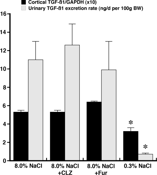
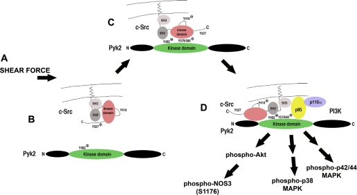
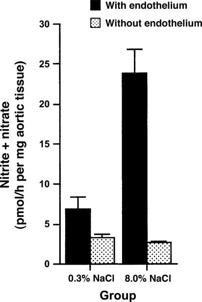
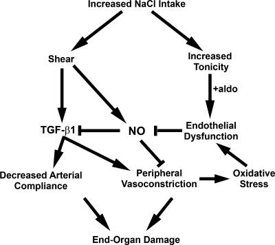
Animal and human studies support an untoward effect of excess dietary NaCl (salt) intake on cardiovascular and renal function and life span. Recent work has promoted the concept that the endothelium, in particular, reacts to changes in dietary salt intake through a complex series of events that are independent of blood pressure and the renin-angiotensin-aldosterone axis. The cellular signaling events culminate in the intravascular production of transforming growth factor-beta (TGF-beta) and nitric oxide in response to increased salt intake. Plasticity of the endothelium is integral in the vascular remodeling consequences associated with excess salt intake, because nitric oxide serves as a negative regulator of TGF-beta production. Impairment of nitric oxide production, such as occurs with endothelial dysfunction in a variety of disease states, results in unopposed excess vascular TGF-beta production, which promotes reduced vascular compliance and augmented peripheral arterial constriction and hypertension. Persistent alterations in vascular function promote the increase in cardiovascular events and reductions in renal function that reduce life span during increased salt intake.


Figures

Fig. 1.
Fig. 1.
Survival of 679 male rats fed formulated diets containing varying amounts of NaCl. Life span was progressively reduced as dietary salt content increased beyond 2.0% NaCl. [Reproduced from Meneely and Ball (38), with permission from Elsevier.]
Fig. 2.
Fig. 2.
Effect of addition of chlorothiazide (CLZ) and furosemide (Fur) on renal cortical expression of transforming growth factor (TGF)-β1, relative to GAPDH, and urinary excretion of TGF-β1 in rats fed 8.0% NaCl diet for 4 days. Neither diuretic altered these parameters compared with data obtained from rats maintained on the high-salt diet, suggesting that diuretics are ineffective in reducing salt-induced intrarenal production of TGF-β1. *P < 0.05 vs. other groups. [Data obtained from Ying and Sanders (69).]
Fig. 3.
Fig. 3.
Working model of salt-induced endothelial cell activation. A: introduction of shear force activates the endothelium by opening a tetraethylammonium-inhibitable potassium channel. B: Pyk2 is autophosphorylated at Y402 and activated. C: c-Src is recruited and activated by Pyk2. Activity of Pyk2 is increased by c-Src-mediated phosphorylation at Y579/580 in the kinase domain. D: phosphatidylinositol 3-kinase (PI3K) is recruited to the complex, permitting activation of Akt and calcium-independent activation of nitric oxide synthase isoform 3 (NOS3) by phosphorylation at S1176 in rats. This complex also activates p38 and p42/44 MAPK pathways, resulting in augmented endothelial cell production of TGF-β1.
Fig. 4.
Fig. 4.
Aortic ring sections from Sprague-Dawley rats fed 0.3% NaCl diet produced less nitrite, a metabolite of nitric oxide (NO), than ring sections from Sprague-Dawley rats fed 8.0% NaCl diet. Effect was lost with removal of the endothelium. [Data obtained from Ying and Sanders (67).]
Fig. 5.
Fig. 5.
Proposed mechanistic link between dietary salt intake and end-organ damage mediated through alteration in vascular structure and function. Excess dietary salt intake induces endothelial shear forces that increase production of TGF-β1 and NO (67, 69, 72, 73). In the setting of salt-induced increased tonicity and aldosterone (aldo), endothelial cells in culture become dysfunctional and produce less NO (41). Oxidative stress, induced by hypertension associated with peripheral vasoconstriction or by other disease states (2, 25, 34, 50), also promotes endothelial dysfunction and diminished NO production. These factors become critical determinants of outcome, since NO serves a vasodilator function and inhibits endothelial TGF-β1 production (71). Fibrogenic effects of TGF-β1 promote a decrease in arterial compliance, and the role of TGF-β1 as a peripheral vasoconstrictor facilitates hypertension (75). Both processes contribute to end-organ damage.

References

    1. Akagi Y, Isaka Y, Arai M, Kaneko T, Takenaka M, Moriyama T, Kaneda Y, Ando A, Orita Y, Kamada T, Ueda N, Imai E. Inhibition of TGF-β1 expression by antisense oligonucleotides suppressed extracellular matrix accumulation in experimental glomerulonephritis. Kidney Int 50: 148–155, 1996. - PubMed
    1. Alp NJ, Channon KM. Regulation of endothelial nitric oxide synthase by tetrahydrobiopterin in vascular disease. Arterioscler Thromb Vasc Biol 24: 413–420, 2004. - PubMed
    1. Annes JP, Munger JS, Rifkin DB. Making sense of latent TGFβ activation. J Cell Sci 116: 217–224, 2003. - PubMed
    1. Avraham H, Park SY, Schinkmann K, Avraham S. RAFTK/Pyk2-mediated cellular signalling. Cell Signal 12: 123–133, 2000. - PubMed
    1. Azuma N, Akasaka N, Kito H, Ikeda M, Gahtan V, Sasajima T, Sumpio BE. Role of p38 MAP kinase in endothelial cell alignment induced by fluid shear stress. Am J Physiol Heart Circ Physiol 280: H189–H197, 2001. - PubMed

Publication types

MeSH terms

Substances