. 2009 Jul;81(1):222-30.
doi: 10.1095/biolreprod.108.074666. Epub 2009 Apr 1.

Manipulating actin dynamics affects human in vitro decidualization

Affiliations

Manipulating actin dynamics affects human in vitro decidualization

Ivanna Ihnatovych et al. Biol Reprod. 2009 Jul.

Abstract

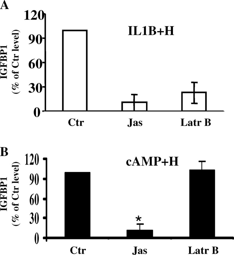
The differentiation of uterine stromal fibroblasts into decidual cells is critical for establishing pregnancy. This process, called decidualization, requires the reorganization of the actin cytoskeleton, which mainly depends on actin dynamics and the phosphorylation status of the myosin light chain. We manipulated actin dynamics with jasplakinolide (100 nM) and latrunculin B (1 microM), both of which significantly inhibited the synthesis of decidualization markers induced by 6 days of treatment with embryo-mimicking stimulus interleukin 1beta (IL1B) and steroid hormones (SHs; 17beta-estradiol and medroxyprogesterone acetate) in the human uterine fibroblast (HuF) in vitro model. However, only jasplakinolide had long-lasting effects on the G-actin:F-actin ratio and prevented decidualization induced by the artificial stimulus cAMP (and SHs). Actin-binding protein cofilin mainly colocalized with G-actin in the nucleus as well as the cytoplasm. Only some spots of colocalization between cofilin and F-actin were detected in the cytoplasm. Brief extraction of cytosolic proteins from living cells revealed that in cells treated with IL1B or cAMP (and SHs) for 6 days, cofilin was mainly detected in the nucleus. The translocation of cofilin from cytosol to nucleus was also detected in HuFs treated for 12 days with SHs, IL1B and SHs, and cAMP and SHs. The same significant translocation was confirmed in primary baboon stromal uterine fibroblasts. We conclude that changes in actin dynamics, particularly the stabilization of F-actin, have a significant negative impact on decidualization, and the translocation of cofilin to the nucleus is a key feature of this process in the primate.


Figures

FIG. 1.
FIG. 1.
Effect of actin dynamics drug manipulation on decidualization. Human uterine fibroblast cells were pretreated for 1 h with latrunculin B (Latr B; 1 μM), jasplakinolide (Jas; 100 nM), or vehicle control (Ctr), followed by treatment for 6 days with (A) IL1B and hormones (IL1B + H) or (B) cAMP and hormones (cAMP + H). The IGFBP1 protein released into the medium was measured using ELISA. Note that although pretreatment with both inhibitors caused a significant decrease in the synthesis of IGFBP1 in HuFs undergoing decidualization induced by IL1B and hormones (A), only jasplakinolide decreased the level of IGFBP1 in HuFs treated with cAMP and hormones (B) compared with HuFs with vehicle control pretreatment (Ctr). *P < 0.05.
FIG. 2.
FIG. 2.
Effect of latrunculin B and jasplakinolide on actin dynamics in HuF cells. A) The densitometric evaluation of the G-actin:F-actin ratio (labeled as G-/F-actin ratio) in HuF cells pretreated with latrunculin B (Latr B; 1 μM) or jasplakinolide (Jas; 100 nM) and either harvested immediately after 1 h of treatment with the drugs (1 h; white bars) or collected after 1 h of treatment with the drugs followed by incubation with 2% SFBS-RPMI for 24 h (1 h/24 h; black bars). The G-actin:F-actin ratio in untreated cells (Ctr) was set as 1, and other treatments were compared with the control. The blot at the top shows representative immunoblots of the distribution of beta-actin (ACTB) in the G-actin and F-actin fractions of HuF cells detected after 1 h of treatment with the drugs (1 h; upper blot) and after 1 h of treatment with the drugs followed by incubation with 2% SFBS-RPMI for 24 h (1 h/24 h; lower blot). B) The densitometric evaluation of the G-actin:F-actin ratio (labeled as G-/F-actin ratio) in HuF cells pretreated for 1 h with jasplakinolide (Jas; black bars) or with vehicle control (Ctr; white bars), followed by the treatment for 6 days with hormones (H), IL1B and hormones (IL1B + H), cAMP and hormones (cAMP + H), or untreated controls (Ctr). The blot at the top shows representative immunoblots of the distribution of ACTB in the G-actin and F-actin fractions of HuF cells treated with jasplakinolide (Jas; lower blot) or without pretreatment (upper blot) as detected after 6 days of decidualization treatment of the cells. Note the overall decrease in the G-actin:F-actin ratio detected after 6 days of decidualization treatment in HuF cells pretreated with jasplakinolide compared with HuFs without pretreatment.
FIG. 3.
FIG. 3.
Association of cofilin with monomeric actin. Confocal microscopy of double-immunofluorescent staining for G-actin (visualized with DNAse I Texas Red dye; DNAse I; middle column) and cofilin (green; cofilin; left column) of HuF cells treated with hormones (H), IL1B and hormones (IL1B + H), cAMP and hormones (cAMP + H), or vehicle control (Ctr) for 6 days. Merged image is shown in the right column. All images were taken at the same magnification. Bar = 20 μm.
FIG. 4.
FIG. 4.
Association of cofilin with filamentous actin during cell decidualization and after extraction of cytosolic proteins. A) Confocal microscopy of double-immunofluorescent staining for F-actin (visualized with rhodamine-conjugated phalloidin [red; Phalloidin column]) and cofilin (green; Cofilin column) in vehicle-treated HuF cells (Ctr) and in HuFs treated for 6 days with hormones (H), IL1B and hormones (IL1B + H), or cAMP and hormones (cAMP + H). Images in rightmost column provide higher-magnification detail of areas close to arrowheads on merge images (Detail). Bar = 20 μm. B) Confocal microscopy (after a 1-min extraction of the soluble cytosolic proteins in live cells) of double-immunofluorescent staining for F-actin (visualized with rhodamine-conjugated phalloidin (red; Phalloidin column]) and cofilin (green; Cofilin column) in vehicle-treated HuF cells (Ctr) and in HuFs treated for 6 days with hormones (H), IL1B and hormones (IL1B + H), or cAMP and hormones (cAMP + H). Images in rightmost column (Detail) provide higher-magnification detail. Arrows in details indicate cofilin-phalloidin colocalization. Bar = 20 μm.
FIG. 5.
FIG. 5.
Changes in phosphorylation and localization of cofilin during decidualization. A) Distribution of p-cofilin and cofilin in the cytosolic (c) and nuclear extract (n) fractions of HuFs treated for 6 days with hormones (H), IL1B and hormones (IL1B + H), cAMP and hormones (cAMP + H), and vehicle controls (Ctr) as detected by Western blotting with specific antibodies. The cytosolic:nuclear fraction ratio in untreated cells was set as 1, and other treatments were compared with the control. B) The densitometric evaluation of the cytosol:nuclear extract ratio (labeled as Cyt./Nucl. extr) for p-cofilin (pCofilin; upper graph; white bars) and cofilin (lower graph; black bars) in HuFs treated for 6 days with hormones (H), IL1B and hormones (IL1B + H), cAMP and hormones (cAMP + H), and vehicle-treated controls (Ctr). The ratio in untreated cells was set as 1, and other treatments were compared with the control. Note the significant decrease (*P < 0.05) in the cytosol:nucleus ratio for cofilin in HuFs treated for 6 days with cAMP and hormones in comparison with Ctr.
FIG. 6.
FIG. 6.
Localization of cofilin in HuF cells treated with decidualization stimuli. A and B) Confocal microscopy of immunofluorescent staining of cofilin (green; left columns) in vehicle-treated HuF cells (Ctr) and in HuFs undergoing decidualization for 6 days (A) and 12 days (B) induced by hormones (H), IL1B and hormones (IL1B + H), and cAMP and hormones (cAMP + H). Nuclear staining with DAPI (blue; middle columns) and merged images (right columns) are shown. Bar = 20 μm. C) The densitometric evaluation of the ratio of immunofluorescent staining of cofilin in the cytosol to immunofluorescent staining of cofilin in the nucleus in vehicle-treated HuF cells (Ctr) and in HuFs undergoing decidualization for 6 days (white bars) and 12 days (black bars) induced by hormones (H), IL1B and hormones (IL1B + H), and cAMP and hormones (cAMP + H). Note the significant decrease (*P < 0.05) in the cytosol:nucleus ratio (labeled as Cyt./Nucl.) for cofilin staining in HuFs treated for 6 days with cAMP and hormones in comparison with control, and for cofilin staining in all 12-day decidualization-inducing treatments in comparison with control.
FIG. 7.
FIG. 7.
Localization of cofilin in primary baboon uterine stromal cells treated with decidualization stimuli for 12 days. A) Confocal microscopy of immunofluorescent staining of cofilin (green; left column) in vehicle-treated cells (Ctr) and in cells undergoing decidualization induced by hormones (H), IL1B and hormones (IL1B + H), and cAMP and hormones (cAMP + H) for 12 days. Nuclear staining with DAPI (blue; middle column) is shown. Merged images are shown in the right column. Bar = 20 μm. B) The densitometric evaluation of the ratio of immunofluorescent staining of cofilin in the cytosol to immunofluorescent staining of cofilin in the nucleus in control-treated HuF cells (Ctr) and in HuFs undergoing decidualization for 12 days induced by hormones (H), IL1B and hormones (IL1B + H), and cAMP and hormones (cAMP + H). Note the significant decrease (*P < 0.05) in the cytosol:nucleus ratio (labeled as Cyt./Nucl.) for cofilin staining in all decidualization-inducing treatments in comparison with control.

Similar articles

Cited by

References

    1. Ayscough KR, Winder SJ. Two billion years of actin. Meeting on cytoskeletal dynamics: from cell biology to developmental disease. EMBO Rep 2004; 5: 947 952 - PMC - PubMed
    1. Strakova Z, Mavrogianis P, Meng X, Hastings JM, Jackson KS, Cameo P, Brudney A, Knight O, Fazleabas AT. In vivo infusion of interleukin-1β and chorionic gonadotropin induces endometrial changes that mimic early pregnancy events in the baboon. Endocrinology 2005; 146: 4097 4104 - PubMed
    1. Kim JJ, Jaffe RC, Fazleabas AT. Insulin-like growth factor binding protein-1 expression in baboon endometrial stromal cells: regulation by filamentous actin and requirement for de novo protein synthesis. Endocrinology 1999; 140: 997 1004 - PubMed
    1. Ihnatovych I, Hu W, Martin JL, Fazleabas AT, de Lanerolle P, Strakova Z. Increased phosphorylation of myosin light chain prevents in vitro decidualization. Endocrinology 2007; 148: 3176 3184 - PubMed
    1. Paavilainen VO, Bertling E, Falck S, Lappalainen P. Regulation of cytoskeletal dynamics by actin-monomer-binding proteins. Trends Cell Biol 2004; 14: 386 394 - PubMed

Publication types