. 2009 Aug;50(8):3826-32.
doi: 10.1167/iovs.08-2694. Epub 2009 Apr 1.

Cyclic mechanical stress and trabecular meshwork cell contractility

Affiliations

Cyclic mechanical stress and trabecular meshwork cell contractility

Renata F Ramos et al. Invest Ophthalmol Vis Sci. 2009 Aug.

Abstract

Purpose: Ocular pulse decreases outflow facility of perfused anterior segments. However, the mechanism by which conventional outflow tissues respond to cyclic intraocular pressure oscillations is unknown. The purpose of the present study was to examine responses of trabecular meshwork (TM) cells to cyclic biomechanical stress in the presence and absence of compounds known to affect cell contractility.

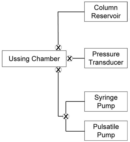
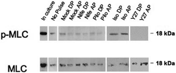
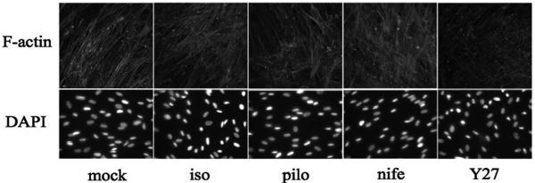
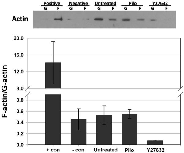
Methods: To model flow in the juxtacanalicular region of the TM and to measure changes in transendothelial flow, human TM cell monolayers on permeable filters were perfused at a constant flow rate until reaching a stable baseline pressure and then were exposed to cyclic stress with an average amplitude of 2.7 mm Hg peak to peak at a 1-Hz frequency for 2 hours in the presence or absence of compounds known to affect cell contractility (isoproterenol, Y27632, pilocarpine, and nifedipine). Pressure was recorded continuously. Immunocytochemistry staining was used to determine filamentous actin stress fiber content, whereas Western blot analysis was used to measure the extent of myosin light chain (p-MLC) phosphorylation and ratio of filamentous to globular actin.

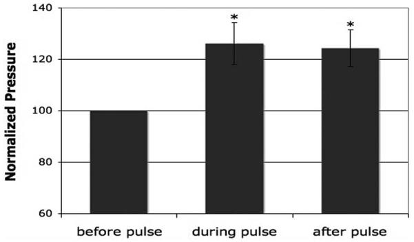
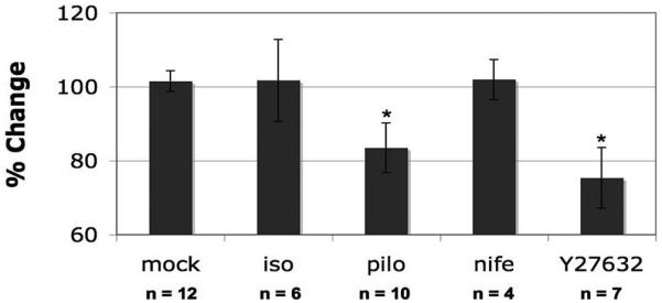
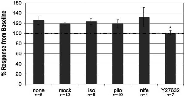
Results: Human TM cells respond to cyclic pressure oscillations by increasing mean intrachamber pressure (decreasing hydraulic conductivity) (126.13% +/- 2.4%; P < 0.05), a response blocked in the presence of Y27632, a rho-kinase inhibitor (101.35 +/- 0.59; P = 0.234), but not isoproterenol, pilocarpine, or nifedipine. Although mechanical stress appeared to have no effect, Y27632 decreased phosphorylated myosin light chain, filamentous/globular actin ratio, and stress fiber formation in TM cells.

Conclusions: Human TM cells respond to cyclic mechanical stress by increasing intrachamber pressure. Pulse-mediated effects are blocked by Y27632, implicating a role for Rho-kinase-mediated signaling and cellular contractility in ocular pulse-associated changes in outflow facility.


Figures

Figure 1
Figure 1
Simplified schematic diagram of the constant flow cell perfusion system modified to introduce intrachamber pressure oscillations. Transcellular flow in Ussing chamber occurred in an apical-to-basal direction.
Figure 2
Figure 2
Summary of mean intrachamber pressures of perfused human TM monolayers before, during, and after the introduction of cyclic pressure oscillations. A significant increase in pressure was observed in the presence of cyclic biomechanical stress and was maintained after the pressure oscillations were stopped (*P ≤ 0.05; n = 6). Average perfusion time was 5.29 ± 0.93 hours. Results are presented as mean ± SEM.
Figure 3
Figure 3
Percentage change in intrachamber pressure in response to drug treatment of human TM monolayers. TM cells were perfused under constant flow. Pressure was recorded and compared before and after the exchange of chamber contents for DMEM (control), isoproterenol (iso), pilocarpine (pilo), nifedipine (nife), or Y27632 (*P ≤ 0.05). Total duration of the perfusion was 10.97 ± 1.13 hours for the control group, 10.11 ± 0.66 hours for the isoproterenol group, 10.5 ± 0.96 hours for the pilocarpine group, 8.71 ± 2.37 hours for the nifedipine group, and 10.14 ± 1.24 hours for the Y27632 group. Results are shown as mean ± SEM.
Figure 4
Figure 4
Effect of cyclic pressure oscillations in perfused human TM monolayers in the presence of different compounds. Baseline for each perfusion was normalized to the intrachamber pressure before cyclic biomechanical stress was introduced (*P ≤ 0.01). Total durations of the perfusion in hours were 10.97 ± 1.13 for the control group, 10.11 ± 0.66 for isoproterenol, 10.5 ± 0.96 for pilocarpine, 8.71 ± 2.37 for nifedipine, and 10.14 ± 1.24 for the Y27632 group. Results are shown as mean ± SEM.
Figure 5
Figure 5
Changes in MLC phosphorylation in perfused human TM monolayer exposed to pulse. Analysis of cell lysates collected from TM monolayers grown on polycarbonate filters before perfusion (in culture or no pulse) or after perfusion. Shown are the results of one representative experiment of two total experiments for each time point in each treatment group. Top: protein levels of phosphorylated myosin light chain (p-MLC). Bottom: levels of total myosin light chain (MLC). Samples were collected after a chamber exchange with only DMEM (mock) or treated DMEM. nife, nifedipine; pilo, pilocarpine; iso, isoproterenol; Y27, Y27632; DP, during pulse; AP, after pulse.
Figure 6
Figure 6
Visualization of human TM cells on coverslips stained for F-actin (confocal microscopy) and DAPI-stained nuclei (epifluorescence microscopy). Analysis of F-actin stress fiber formation of TM cells in response to 1-hour treatments with DMEM (mock), isoproterenol (iso), pilocarpine (pilo), nifedipine (nife), or Y27632 (Y27). TM cells used for immunostaining showed similar levels of monolayer confluence between treatment groups. Results display representative experiments from a total of three experiments for each treatment group.
Figure 7
Figure 7
Effects of Y27632 and pilocarpine on actin polymerization in TM cell monolayers. The proportion of filamentous (F)-actin to globular (G)-actin in TM cells was analyzed after 30-minute treatments with 10 μM pilocarpine (pilo), 10 μM Y27632, 1 μM phalloidin (+ con), 10 μM cytochalasin (- con) or an untreated control. Top: actin distribution obtained by immunoblot analysis in one experiment. Bottom: summary of results (mean ± SEM) obtained from three combined experiments.

References

    1. Hashimoto JM, Epstein DL. Influence of intraocular pressure on aqueous outflow facility in enucleated eyes of different mammals. Invest Ophthalmol Vis Sci. 1980;19:1483–1489. - PubMed
    1. Johnstone MA. Pressure-dependent changes in nuclei and the process origins of the endothelial cells lining Schlemm’s canal. Invest Ophthalmol Vis Sci. 1979;18:44–51. - PubMed
    1. Gonzalez P, Epstein DL, Borras T. Genes upregulated in the human trabecular meshwork in response to elevated intraocular pressure. Invest Ophthalmol Vis Sci. 2000;41:352–361. - PubMed
    1. Borras T, Rowlette LL, Tamm ER, Gottanka J, Epstein DL. Effects of elevated intraocular pressure on outflow facility and TIGR/MYOC expression in perfused human anterior segments. Invest Ophthalmol Vis Sci. 2002;43:33–40. - PubMed
    1. Bradley JM, Kelley MJ, Rose A, Acott TS. Signaling pathways used in trabecular matrix metalloproteinase response to mechanical stretch. Invest Ophthalmol Vis Sci. 2003;44:5174–5181. - PubMed

Publication types

MeSH terms