

Review
doi: 10.1172/JCI37335.
Epub 2009 Apr 1.
Schizophrenia from a neural circuitry perspective: advancing toward rational pharmacological therapies
Affiliations
- PMID: 19339762
- PMCID: PMC2662560
- DOI: 10.1172/JCI37335
Item in Clipboard
Review
Schizophrenia from a neural circuitry perspective: advancing toward rational pharmacological therapies
J Clin Invest.
2009 Apr.
Abstract
Schizophrenia is a severe disorder that disrupts the function of multiple brain systems, resulting in impaired social and occupational functioning. The etiology and pathogenesis of schizophrenia appear to involve the interplay of a potentially large number of genetic liabilities and adverse environmental events that disrupt brain developmental pathways. In this Review, we discuss a strategy for determining how particular common and core clinical features of the illness are associated with pathophysiology in certain circuits of the cerebral cortex. The identification of molecular alterations in these circuits is providing critical insights for the rational development of new therapeutic interventions.
Figures

According to this view, the etiology or cause of schizophrenia unleashes pathogenetic mechanisms that produce specific pathological entities. Each of these conserved sets of molecular and cellular disturbances in the brain alters the normal circuitry and function of the brain so that the resulting pathophysiology gives rise to distinct components of the clinical syndrome recognized as schizophrenia. The bidirectional arrows indicate the following: (a) understanding pathophysiology and pathogenesis is required for the rational identification of novel molecular targets for improving treatment and secondary prevention, respectively, and (b) proof-of-concept validation of compounds with activity at these targets requires normalization of biomarkers for these processes.
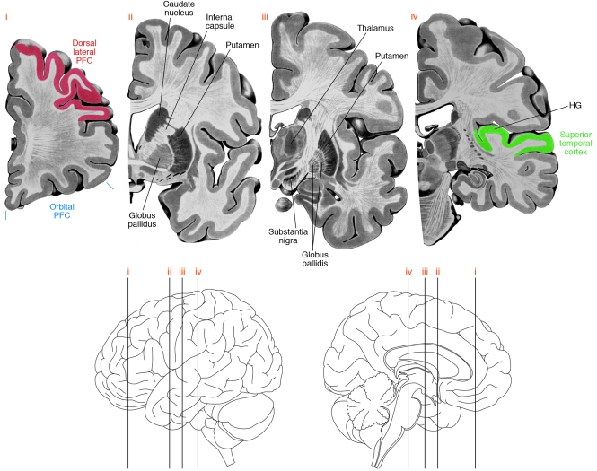
Four coronal sections (top) through the left hemisphere of the human brain at the approximate levels shown in the lateral and medial views (bottom). Some of the brain regions implicated in neural circuitry disturbances in schizophrenia are indicated. Note that the AI is located within HG. PFC, prefrontal cortex.
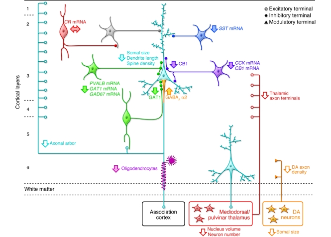
Pyramidal neurons (light blue) in deep layer 3 have smaller somal size, shorter basilar dendrites, lower dendritic spine density, and a reduced axonal arbor in schizophrenia. Altered GABA neurotransmission by PVALB-containing neurons (green) is indicated by expression deficits in several gene products as well as by lower levels of GAT1 protein in the terminals of chandelier neurons and upregulated GABAA receptor α2 subunits at their synaptic targets, the axon initial segments of pyramidal neurons. Expression of the neuropeptide somatostatin (SST) is decreased in GABA neurons (dark blue) that target the distal dendrites of pyramidal neurons. Decreased cholecystokinin (CCK) and cannabinoid receptor 1 (CB1) mRNA levels and lower CB1 protein in axon terminals suggest altered regulation of GABA neurotransmission in a subset of basket neurons (purple) that target the cell body and proximal dendrites of pyramidal neurons. Gene expression does not seem to be altered in calretinin-containing (CR-containing) GABA neurons (red) that primarily target other GABA neurons (gray). Putative alterations in thalamic and DA cell bodies and their projections to the DLPFC are also shown. Some studies indicate that the number and/or gene expression in oligodendrocytes is also altered (119). Not all of the circuitry alterations shown here have been sufficiently replicated or demonstrated to be specific to the disease process of schizophrenia to be considered established facts; filled arrows indicate abnormalities supported by convergent and/or replicated observations. Figure adapted with permission from Neuron (1).
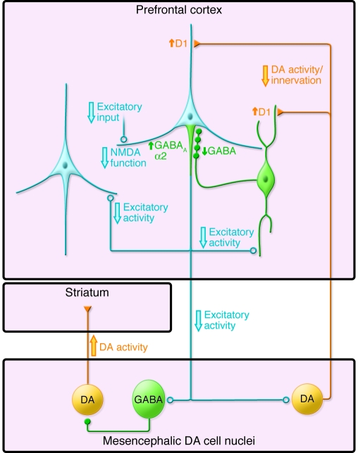
In one view, the excitatory activity of cortical pyramidal neurons (light blue) is thought to be reduced in schizophrenia due to lower excitatory inputs, NMDA receptor hypofunction, and/or decreased DA neurotransmission through D1 receptors. Similar alterations may contribute to reduced GABA synthesis in PVALB chandelier (green) neurons and a compensatory increase in GABAA receptors containing α2 subunits in the axon initial segment of pyramidal neurons. Decreased excitatory input to neurons in the mesencephalon would lead to increased DA activity in the striatum and decreased DA activity in the cortex, with compensatory but functionally insufficient upregulation of D1 receptors. See text for additional details. Figure adapted with permission from Nature Medicine (125).
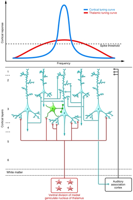
Auditory cortical processing is initiated by projections from the medial geniculate nucleus of the thalamus. These projections are arranged tonotopically (i.e., along a frequency gradient that is broadly tuned). The subsequent activation of a reciprocally connected isofrequency network of pyramidal cells (light blue) within layer 3 selectively amplifies a narrower preferred frequency, refining the thalamic tuning curve. Densities of dendritic spines and axonal boutons are reduced in deep layer 3 of subjects with schizophrenia, potentially limiting activation and current flow in the pyramidal cell network. Pyramidal neurons are co-tuned (i.e., they receive concurrent stimulation from thalamic or cortical projection neurons) with local inhibitory neurons (green), leading to a stereotyped excitatory-inhibitory sequence of postsynaptic potentials (123), which increases the temporal precision of depolarization and enhances phasic activity of the pyramidal neuron network (124). Thus, though alterations in GABA neurons in the AI (as described in DLPFC of subjects with schizophrenia; see Figure 2) are currently understudied, if conserved across regions (65), they may further contribute to impaired activation of isofrequency pyramidal neuron networks.
References
-
- Parks, J., Svendsen, D., Singer, P., and Foti, M. 2006. Morbidity and mortality in people with serious mental illness. National Association of State Mental Health Program Directors (NASMHPD) Medical Directors Council. Alexandria, Virginia, USA. http://www.bu.edu/cpr/resources/wellness-summit/documents/morbidity-and-....
-
- Green M.F. What are the functional consequences of neurocognitive deficits in schizophrenia? Am. J. Psychiatry. 1996;153:321–330. - PubMed
