

Review
doi: 10.1172/JCI37703.
Bipolar disorder: from genes to behavior pathways
Affiliations
- PMID: 19339764
- PMCID: PMC2662565
- DOI: 10.1172/JCI37703
Item in Clipboard
Review
Bipolar disorder: from genes to behavior pathways
J Clin Invest.
2009 Apr.
Abstract
Bipolar disorder (BPD) is a devastating illness that is characterized by recurrent episodes of mania and depression. In addition to these cyclic episodes, individuals with BPD exhibit changes in psychovegetative function, cognitive performance, and general health and well being. In this article we draw from neuroimaging findings in humans, postmortem data, and human genetic and pharmacological studies as well as data from animal models of behavior to discuss the neurobiology of BPD. We conclude with a synthesis of where the field stands and with suggestions and strategies for future areas of study to further increase our conceptual understanding of this complex illness.
Figures
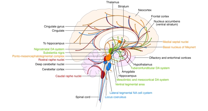
Nuclei as well as their projections are color coded: yellow, cholinergic; green,
dopaminergic; blue, noradrenergic; red, serotonergic.
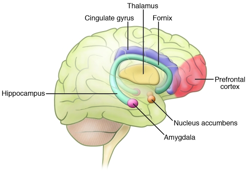
Neuroimaging studies, observations from patients with selective CNS lesions, as
well as data from animal behavioral studies have implicated several regions
throughout the brain in the control of mood states and emotions. These regions are
located throughout the limbic, striatal, and frontal regions. Adapted with
permission from Neuropsychopharmacology (24).
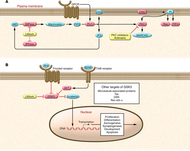
(A) Lithium directly inhibits key enzymes including IPPase and IMPase
that regulate inositol-1,4,5-triphosphate (IP3) recycling.
IMPase–mediated catalysis represents the final step in the conversion
of inositol monophosphate (IMP) to myoinositol. Thus, inhibiting IMPase can reduce
myoinositol levels. The inositol depletion hypothesis proposes that IMPase
inhibition interferes with PI synthesis. The PI signaling cascade starts with
surface receptor activation, shown here as GPCR-mediated activation of PLC.
Activated PLC catalyzes the hydrolysis of PI biphosphate (PIP2) to diacylglycerol
(DAG) and IP3. DAG activates PKC, which, among many other functions, activates
myristoylated, alanine-rich C-kinase substrate (MARCKS). The antimanic drugs
lithium and VPA both decrease levels of phosphorylated and total MARCKS. Molecules
in red depict enzymes, while molecules in blue depict second messengers. PI
signaling modulates other second messenger proteins including the small GTPase,
Ras, and phosphatidylinositol-4-phosphate (PIP4). (B) Overview of the
Wnt and GSK3 signaling pathways. Both lithium and Wnt signaling can inhibit GSK3.
In the Wnt signaling pathway, Wnt glycoproteins interact with the frizzled family
of receptors to stimulate the dishevelled-mediated (not shown) inactivation of
GSK3. Inhibition of GSK3 prevents β-catenin phosphorylation, which
inhibits its degradation and allows it to act as a transcription factor. Wnt
proteins have been implicated in the regulation of neuron morphology,
neurotransmission, and synaptogenesis. GSK3 is also inhibited by AKT (also known
as PKB), which is activated downstream of the tropomyosin receptor B (TrkB), whose
ligand is BDNF. In addition to inhibiting β-catenin, GSK3 has numerous
other targets including microtubule-associated proteins, tau, GSK, and rev-erb
α. Adapted with permission from
Neuropsychopharmacology (24).
References
-
- Akiskal H.S., et al. Re-evaluating the prevalence of and diagnostic composition within the broad clinical spectrum of bipolar disorders. . J. Affect. Disord. 2000;59(Suppl. 1):S5–S30. - PubMed
Publication types
MeSH terms
LinkOut - more resources
Full Text Sources
Medical
Molecular Biology Databases
