Review
. 2009 Mar;4(2):88-95.
doi: 10.1097/COH.0b013e3283223d8d.

Chemokine receptors and other G protein-coupled receptors

Affiliations
Review

Chemokine receptors and other G protein-coupled receptors

David T Lodowski et al. Curr Opin HIV AIDS. 2009 Mar.

Abstract

Purpose of review: Class A G protein-coupled receptors (GPCRs), including the chemokine receptors, CCR5 and CXCR4, share a seven transmembrane-spanning alpha-helix architecture that accommodates signal propagation from across biological membranes. CXCR4 and CCR5 are utilized as co-receptors during HIV viral entry and, therefore, crystal structures of GPCRs aid in the understanding of their function in viral entry.

Recent findings: Recent progress in structure determination of class A GPCRs, which include vertebrate and invertebrate rhodopsin as well as adrenergic and adenosine receptors, provides molecular templates for how this diverse group of transmembrane receptors functions. Each of these GPCRs differs in how specific ligands bind to the transmembrane core, underscoring that additional structures of GPCRs from other subfamilies are needed to facilitate rational drug design. More recent studies also indicate a need to consider the more complex character of GPCRs, such as their oligomerization and dynamics.

Summary: Recently, the atomic structures of invertebrate rhodopsin, beta1-adrenergic and beta2-adrenergic receptors and the A(2A)-adenosine receptor have been solved via X-ray crystallography. The impact that these structures have on the biochemistry of viral entry and signal transduction is discussed. Because the chemokine receptors have proven refractory to structural studies thus far, further structural study of the chemokine receptors will be essential to understanding ligand binding, activation and function as co-receptors during viral entry.


Figures

Figure 1
Figure 1
Schematic representation of how GPCRs function in A. signal transduction and B. HIV-1 viral entry. A. The GPCR: heterotrimeric G protein signaling mechanism. Upon agonist binding to the receptor, the GPCR undergoes a structural change, freeing it to form a ternary complex with a heterotrimeric G protein. Formation of the ternary complex induces the release of the bound GDP from the α subunit and a GTP molecule binds to the empty nucleotide binding site. This event causes the α and βγ subunits to dissociate and each is free to then bind to cellular effector enzymes such as adenylyl cyclase or phospholipases. These effectors produce second messenger compounds which activate downstream signaling proteins, ultimately resulting in a cellular response. Hydrolysis of the bound GTP on the α subunit results in inactivation and this GDP bound α binds to a free βγ subunit, thus reforming the inactive heterotrimer. B. GPCRs as co-receptors for viral entry. Trimers of the Env protein are expressed upon the surface of the viral envelope. The gp120 subunit of Env binds to the CD4 cellular receptor on a leukocyte. This event serves to tether the virus to the membrane, and induces structural changes within gp120 which make it possible to bind to the chemokine (CXCR4 or CCR5) GPCRs. Upon GPCR binding, the gp41 subunit undergoes structural changes and inserts helices into the membrane, initializing fusion of the viral membrane with the cellular membrane.
Figure 1
Figure 1
Schematic representation of how GPCRs function in A. signal transduction and B. HIV-1 viral entry. A. The GPCR: heterotrimeric G protein signaling mechanism. Upon agonist binding to the receptor, the GPCR undergoes a structural change, freeing it to form a ternary complex with a heterotrimeric G protein. Formation of the ternary complex induces the release of the bound GDP from the α subunit and a GTP molecule binds to the empty nucleotide binding site. This event causes the α and βγ subunits to dissociate and each is free to then bind to cellular effector enzymes such as adenylyl cyclase or phospholipases. These effectors produce second messenger compounds which activate downstream signaling proteins, ultimately resulting in a cellular response. Hydrolysis of the bound GTP on the α subunit results in inactivation and this GDP bound α binds to a free βγ subunit, thus reforming the inactive heterotrimer. B. GPCRs as co-receptors for viral entry. Trimers of the Env protein are expressed upon the surface of the viral envelope. The gp120 subunit of Env binds to the CD4 cellular receptor on a leukocyte. This event serves to tether the virus to the membrane, and induces structural changes within gp120 which make it possible to bind to the chemokine (CXCR4 or CCR5) GPCRs. Upon GPCR binding, the gp41 subunit undergoes structural changes and inserts helices into the membrane, initializing fusion of the viral membrane with the cellular membrane.
Figure 2
Figure 2
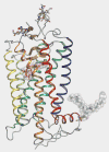
Atomic structure of rhodopsin, a prototypical GPCR. Helices are colored according to their primary sequence: helix-I, blue; helix-II, blue-green, helix-III, green; helix-IV, lime-green; helix-V, yellow; helix-VI, orange; helix-VII, red; helix-8, purple. Chromophore is colored as transparent hot-pink surface. The initial crystal structure revealed the topology of these individual helices and the conformation of the chromophore allowing a structural understanding of previously determined biochemical and biophysical studies of rhodopsin. Later improvements in resolution revealed the presence of ordered waters within the transmembrane region as well as the complete polypeptide chain. The region above the receptor is the extracellular face and below is the cytoplasmic (G protein interacting) face (PDB ID:1U19).
Figure 3
Figure 3
Representative structures of several GPCRs. All structures are in either the ground (inactive) state or have inverse agonist bound. A. The 2.6 Å structure of invertebrate (squid) rhodopsin (PDB ID:2Z73). B. The 2.8 Å structure of the β1-adrenergic receptor (PDB ID:2VT4). C. The 2.4 Å structure of the T4 lysozyme β2-adrenergic receptor fusion (PDB ID:2RH1). D. The 2.6 Å structure of the T4 lysozyme-A2A adenosine receptor (PDB ID:3EML). While each of these receptors has evolved to bind different ligands and G proteins, the overall seven transmembrane helix architecture and topology has been retained. All GPCRs are shown in the same orientation as rhodopsin in figure 1.
Figure 4
Figure 4
Structural similarities between ground state rhodopsin and the β2-adrenergic receptor. Rhodopsin is represented depicted in red and the β2-adrenergic receptor depicted in light grey. The T4 lysozyme fusion has been removed from β2- adrenergic receptor for clarity. The high degree of structural similarity within the transmembrane regions of the two receptors is very high and root-mean-squared deviations this region are only 3.3 Å. (Panels A & B) Two views of the receptors as viewed in the plane of the plasma membrane. The cytoplasmic loops (C) follow different routes in each receptor and the C-terminal tail is not observed in the adrenergic receptor structure. The extracellular face (D) is also significantly different as this face of the receptor has different functions in each receptor; a diffusible hydrophilic ligand must enter adrenergic receptor whereas the region surrounding the chromophore has evolved to shield it from the extracellular environment.

References

    1. Takeda S, Kadowaki S, Haga T, Takaesu H, Mitaku S. Identification of G protein-coupled receptor genes from the human genome sequence. FEBS Lett. 2002;520:97–101. - PubMed
    1. Kroeze WK, Sheffler DJ, Roth BL. G-protein-coupled receptors at a glance. J Cell Sci. 2003;116:4867–4869. - PubMed
    1. Oldham WM, Hamm HE. Heterotrimeric G protein activation by G-protein-coupled receptors. Nat Rev Mol Cell Biol. 2008;9:60–71. - PubMed
    1. Filipek S, Krzysko KA, Fotiadis D, Liang Y, Saperstein DA, Engel A, Palczewski K. A concept for G protein activation by G protein-coupled receptor dimers: the transducin/rhodopsin interface. Photochem Photobiol Sci. 2004;3:628–638. - PubMed
    1. Park PS, Lodowski DT, Palczewski K. Activation of g protein-coupled receptors: beyond two-state models and tertiary conformational changes. Annu Rev Pharmacol Toxicol. 2008;48:107–141. - PMC - PubMed

Publication types

Substances