

Review
doi: 10.1038/nrn2632.
The neuro-symphony of stress
Affiliations
- PMID: 19339973
- PMCID: PMC2844123
- DOI: 10.1038/nrn2632
Item in Clipboard
Review
The neuro-symphony of stress
Nat Rev Neurosci.
2009 Jun.
Abstract
The impact of stress on brain function is increasingly recognized. Various substances are released in response to stress and can influence distinct neuronal circuits, but the functional advantages of having such a diversity of stress mediators remain unclear. Individual neurotransmitter, neuropeptide and steroid stress mediators have specific spatial and temporal niches, but these niches also overlap. In addition, the effects of individual mediators on neuronal function and plasticity are integrated, and emerging evidence suggests that there is crosstalk between them. Together, this results in the stress instruments producing an orchestrated 'symphony' that enables fine-tuned responses to diverse challenges.
Figures
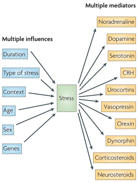
Many factors influence the pattern and magnitude of the response to stress, including the duration of stress exposure (acute versus chronic), the type of stress (physical versus psychological), the stress context (for example, time of day), the developmental stage of the animal (newborn, adult or aged) and the animal's sex and genetic background. The panoply of unique stress situations and neuronal populations that respond to them to affect neural and behavioural plasticity on a timescale from seconds to years is not well served by a single mediator — hence the need for multiple instruments, so that each combination of mediators addresses the specific aspects of a stressor. The molecules that convey the stress signal to the CNS and that contribute to the resulting functional changes in the CNS (stress mediators) include monoamines, neuropeptides and steroid hormones, examples of which are shown on the right. Each mediator has a preferred activity domain in space and time.
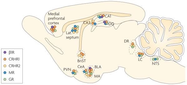
The β1-adrenoceptors for noradrenaline (B1Rs), CRH receptor 1 (CRHR1), CRHR2 and the mineralocorticoid and glucocorticoid receptors (MRs and GRs, respectively) cluster in ‘hot spots’ in the brain. These hot spots include the prefrontal cortex, specific amygdala nuclei, the hippocampus (CA1, CA3 and the dentate gyrus (DG)), the paraventricular nucleus of the hypothalamus (PVN), the dorsal raphe nuclei (DR) and the locus coeruleus (LC). In these areas, receptors for at least two classes of mediators are highly expressed. The hot spots are strategic hubs that connect networks involved in diverse aspects of the brain's stress response, including learning and memory, decision making and hormonal, autonomic and emotional responses. BLA, basolateral amygdala; BnST, bed nucleus of the stria terminalis; CeA, central amygdala; CRH, corticotropin-releasing hormone; lat. septum, lateral septum; MA, medial amygdala; NTS, nucleus tractus solitarii. Data from REFS ,–.
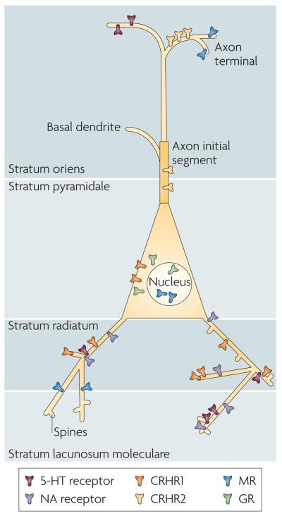
The actions of different stress mediators on a single neuron are orchestrated through the distinct subcellular localization of the relevant receptors. This is illustrated in this cartoon of a hypothetical pyramidal cell (which includes features that exist on either or both CA3 and CA1 principal cells). For example, corticotropin-releasing hormone receptor 1 (CRHR1) is located in somata as well as on dendrites and dendritic spines,,,, whereas CRHR2 seems to reside on the axon initial segment (Y. Chen, personal communication). In addition, monoamine receptors that terminate on hippocampal pyramidal cells reside on specific dendritic domains,. Note that the subcellular distribution patterns of each receptor might differ between brain areas. For instance, glucocorticoid receptors (GRs) might be present on postsynaptic (dendritic) domains in certain amygdala neurons. 5-HT, 5-hydroxytryptamine; MR, mineralocorticoid receptors; NA, noradrenaline.
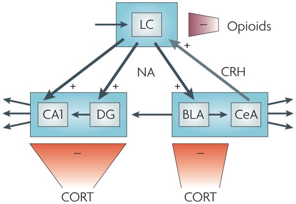
Noradrenaline (NA), corticotropin-releasing hormone (CRH), opioids and corticosterone (CORT) interact in the locus coeruleus (LC) and its projection areas (including the hippocampus and the amygdala) to orchestrate exquisite tuning of neuronal firing patterns in response to stress. Exposure to stress shifts LC noradrenergic cell firing — which is normally kept at a moderate level by glutamatergic input, here represented by the top left arrow — from a moderate tonic activity to high tonic firing that prevents phasic firing. This shift is mediated by CRH projections from the central amygdala on to CRH 1 receptors in the LC. In turn, LC noradrenergic cells project to the basolateral amygdala (BLA), hippocampal CA1 and the dentate gyrus (DG). Here noradrenaline, released shortly after stress exposure, enhances excitability, promoting the encoding of stress-related information. Glutamatergic output from the BLA to the DG is thought to provide a means to ‘emotionally tag’ information processed in the hippocampus, thus rendering it preference in storage. The stress-induced enhancement in activity in the LC, the BLA, the DG and CA1 (which in the DG is aided by rapid corticosteroid actions; see main text) is gradually reversed, resulting in a return to the pre-stress activity level. In the LC, the level of tonic firing is reduced by opiates that bind to κ- and μ-opioid receptors. In the BLA, the DG and CA1, these gradual normalizing effects are exerted by corticosterone, presumably through glucocorticoid receptor-mediated gene-dependent cascades,,. The + signs indicate that the stress mediator enhances cell firing, whereas the–signs indicate decreased cell activity. CeA, central amygdala.
References
-
- De Kloet ER, Joëls M, Holsboer F. Stress and the brain: from adaptation to disease. Nature Rev. Neurosci. 2005;6:463–475. - PubMed
-
- McEwen BS. Physiology and neurobiology of stress and adaptation: central role of the brain. Physiol. Rev. 2007;87:873–904. - PubMed
-
- McGaugh JL. The amygdala modulates the consolidation of memories of emotionally arousing experiences. Annu. Rev. Neurosci. 2004;27:1–28. - PubMed
Publication types
MeSH terms
Substances
Grants and funding
LinkOut - more resources
Full Text Sources
Other Literature Sources
Medical
