. 2009 May 20;28(10):1518-30.
doi: 10.1038/emboj.2009.86. Epub 2009 Apr 2.

Stability of ligand-binding domain dimer assembly controls kainate receptor desensitization

Affiliations

Stability of ligand-binding domain dimer assembly controls kainate receptor desensitization

Charu Chaudhry et al. EMBO J. .

Abstract

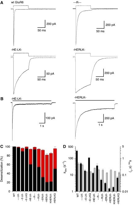
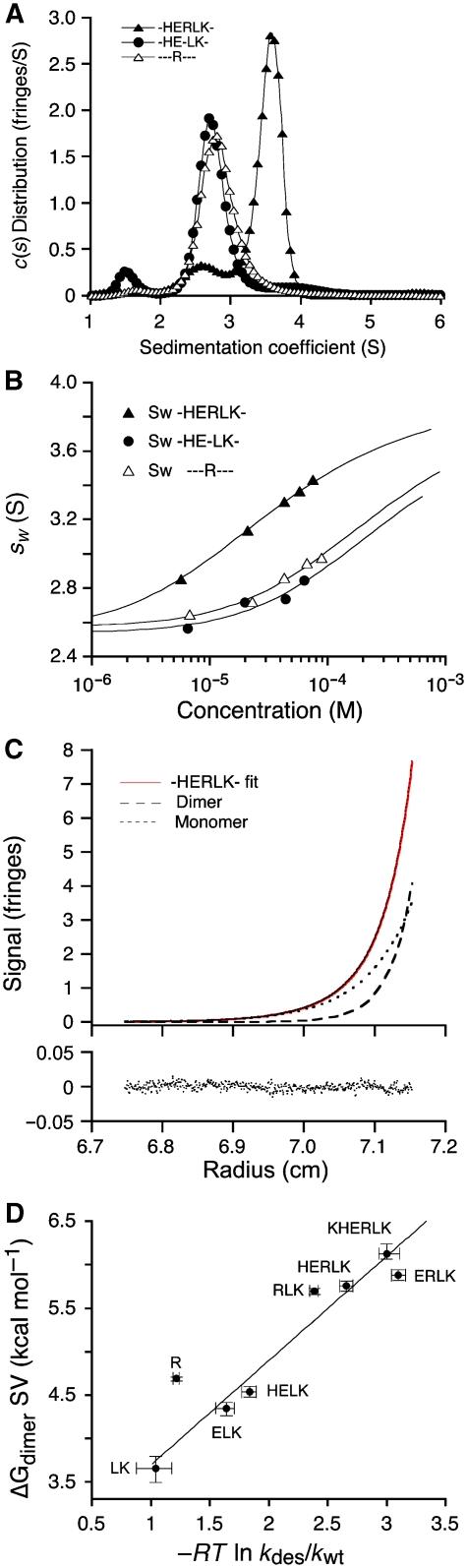
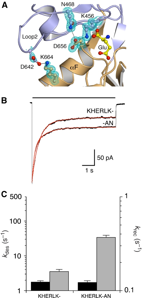
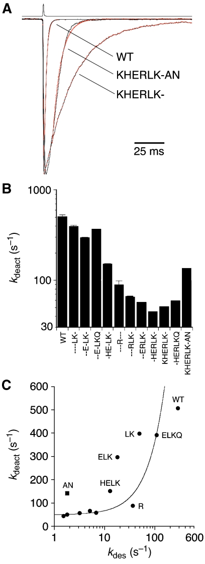
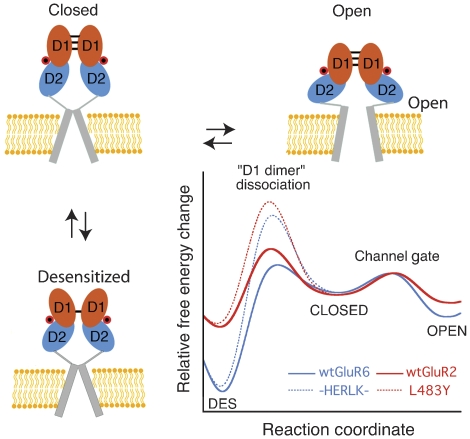
AMPA and kainate receptors mediate fast synaptic transmission. AMPA receptor ligand-binding domains form dimers, which are key functional units controlling ion-channel activation and desensitization. Dimer stability is inversely related to the rate and extent of desensitization. Kainate and AMPA receptors share common structural elements, but functional measurements suggest that subunit assembly and gating differs between these subtypes. To investigate this, we constructed a library of GluR6 kainate receptor mutants and directly measured changes in kainate receptor dimer stability by analytical ultracentrifugation, which, combined with electrophysiological experiments, revealed an inverse correlation between dimer stability and the rate of desensitization. We solved crystal structures for a series of five GluR6 mutants, to understand the molecular mechanisms for dimer stabilization. We demonstrate that the desensitized state of kainate receptors acts as a deep energy well offsetting the stabilizing effects of dimer interface mutants, and that the deactivation of kainate receptor responses is dominated by entry into desensitized states. Our results show how neurotransmitter receptors with similar structures and gating mechanisms can exhibit strikingly different functional properties.


Figures

Figure 1
Figure 1
Conserved clusters of residues differ in the dimer interface of AMPA and kainate receptors. (A) Amino-acid sequence alignment for AMPA and kainate receptor gene families; dimer interface residues exchanged between GluR2 and GluR6 are indicated by Δ; additional residues that play key roles in the effects of individual mutations are indicated by *; cylinders above the alignment indicate location of α-helices B, D, F, and J in GluR6 crystal structures; + indicates the L/Y switch in helix D. (B) Ribbon diagram for wild-type GluR6 shows the location of the critical Tyr490 side chain, surrounded by residues exchanged between GluR2 and GluR6, drawn as gold- and cyan-colored CPK spheres for the pair of subunits in a dimer assembly (a stereo view is shown in Supplementary Figure 1).
Figure 2
Figure 2
Kinetic analysis for a library of GluR6 dimer interface mutants. (A) Responses of outside-out patches to 100-ms applications of 10 mM glutamate are shown for wild type and three mutants. The rate of desensitization (kdes) is fastest for wild type, ∼200-fold slower for -HERLK-, whereas - - -R- - - and -HE-LK- produce intermediate kinetics. (B) Responses to 7-s applications of glutamate show the extent of desensitization for -HE-LK- and -HERLK-, highlighting the impact of the K665R mutation in the -HE-LK- background. (C) The extent of desensitization measured at 100 ms (black) and 7 s (red). (D) Rate of onset of desensitization (black bar) and recovery (grey bar), for wild-type GluR6 and the library of 10 dimer interface mutants; error bars indicate mean±s.e.m.
Figure 3
Figure 3
Dimer formation for GluR6 LBDs measured by analytical ultracentrifugation. (A) c(s) distributions from sedimentation velocity (SV) runs for -HERLK- (1.7 mg ml−1), -HE-LK- (1.9 mg ml−1) and - - -R- - - (2 mg ml−1); concentrations reported in parentheses are derived from peak integration (see also Supplementary Figure 2). Peak positions reflect sedimentation of rapidly interconverting monomer–dimer systems. The ∼3.6 S peak for -HERLK- reflects stronger association of this mutant compared with the 2.8 and 2.9 S peaks for -HE-LK- and - - -R- - -, respectively. (B) Dependence of the weight-average sedimentation coefficient sw on loading concentration for -HERLK-, -HE-LK- and - - -R- - - (symbols), and best fits with a binding isotherm for a monomer–dimer equilibrium (solid lines) with Kd values of 41.2 μM (1σ confidence interval 37–45 μM), 416 μM (1σ 370–460), 321 μM (1σ 307–333) for -HERLK-, -HE-LK- and - - -R- - -, respectively. (C) Representative sedimentation equilibrium profile for -HERLK- derived from a global analysis of data at a range of loading concentrations and rotor speeds, using a monomer–dimer model with a Kd of 102 μM (1σ 85–135). The black line indicates the model used to fit the data, red dots indicate experimental measurements, and dashed lines represent the best-fit populations of monomer and dimer; residuals of the fit are shown below the graph. (D) The free energy change for dimerization ΔGdimer plotted against the free energy change for onset of desensitization relative to wild type (−RT ln kdes/kwt) for each mutant (symbol and error bars). The dotted line shows the best fit from total least-squares optimization, suggesting a linear relationship in which stabilization of the dimer assembly slows the rate of onset of desensitization. A full colour version of this figure is available at The EMBO Journal online.
Figure 4
Figure 4
High-resolution crystal structures for GluR6 LBD mutants. (A) Side chains drawn in stick representation are shown for wild-type GluR6, - - -R- - -, - - -RLK-, -HE-LK- and -HERLK- following least-squares superposition of dimer assemblies using D1 Cα coordinates; a stereo view with CPK spheres for -HERLK- is shown in Supplementary Figure 1. (B) σA-weighted 2mFoDFc electron density maps contoured at 1σ for amino-acid side chains surrounding the native Tyr490 residue; for visualization side chains have been rotated from their orientation in the dimers. (C) Interactions between Tyr490 on one subunit with I749L and Q753K on helix J of the dimer partner for wild-type GluR6 (grey) and -HERLK- (gold). The van der Waals radii of Ile749 for wild-type GluR6 show a steric clash (top panel) with those for Tyr490 in the -HERLK- structure, which is eradicated on mutation to a Leu (bottom panel). (D) Interdomain interactions between helices B and J on one subunit and helices D and F on the other for HERLK and wild-type GluR6. Van der Waals interactions (distance <4 Å) are represented by a solid line connecting partner amino acids; hydrogen bonds by arrows pointing in the direction of the hydrogen bond acceptor; electrostatic (cation–π and salt-bridge) interactions by a thick line; water-mediated interactions with a dashed line; grey and gold indicate bonds for wild-type GluR6 and -HERLK-, respectively.
Figure 5
Figure 5
The K665R mutation forms an intermolecular salt bridge linking helix F with helix J. (A) Dimer assembly for -HERLK- showing side chains for K665R, Glu756 and surrounding water molecules with σA-weighted 2mFoDFc electron density maps at 1.5σ; (B) dimer assembly for wild-type GluR6 illustrating formation of an intramolecular contact between Lys665 and Asp462 in the same subunit; note the 55° difference in χ3 for Glu756. Water molecules W1 and W2 are conserved in the two structures, but the native Lys665 side-chain projects into the second network of water molecules present in -HERLK-.
Figure 6
Figure 6
The E757Q mutation destabilizes dimer assembly. (A) Responses to 100-ms applications of 10 mM glutamate for -HERLK- and -HERLKQ illustrating the five-fold faster rate of onset of desensitization due to E757Q substitution. (B) Sedimentation equilibrium scans for -HERLKQ at 1 mg ml−1 and 24 000 r.p.m. analyzed identically as for -HERLK- (Figure 3C); the dimerization Kd decreases ∼6-fold to 628 μM. (C) Base of helix J tilts by 5° in -HERLKQ; dimer assemblies for -HERLKQ (red) and -HERLK- (gold) were superimposed by least-squares using D1 Cα coordinates; the solid line shows a Cα trace illustrating that movement is limited to the base of helix J. (D) Residues at the base of helix J and in the linker leading to helix K are disordered in -HERLKQ. An FoFc omit electron density map contoured at 3.5σ was calculated with residues Glu756–Lys759 omitted from the Fc calculation; stick representation for these residues show main chain alternative conformations for Glu756, E757Q and Gly758, and side-chain alternative conformations for Glu756, E757Q and Lys759.
Figure 7
Figure 7
Destabilization of ligand binding reduces desensitization. (A) Single subunit of -HERLK- showing glutamate bound in a cleft between D1 (blue) and D2 (orange). Interdomain salt bridge between Lys456 (D1) and Asp656 (D2) and also between Lys664 (D1) and Asp462 (D2) shown with σA-weighted 2mFo DFc electron density at 1σ. (B) Responses to 7-s applications of glutamate reveal an increase in steady-state response for KHERLK-AN compared with KHERLK-. (C) Bar plots showing the effect of K456A/D656N on the rate of desensitization (black) and recovery (grey) in KHERLK-.
Figure 8
Figure 8
Deactivation is greatly slowed in weakly desensitizing GluR6 mutants (A) The rate of deactivation for responses to 1-ms applications of glutamate is much slower for KHERLK- than for wild-type GluR6, 51 and 506 s−1, respectively, but increases for KHERLK-AN to 142 s−1. (B) Bar plots showing the deactivation rate for the library of dimer interface mutants. (C) The rate of deactivation saturates for mutants with stable dimer interfaces; data points for individual mutants are labelled except those for - - -RLK-, - -ERLK-, -HERLK-, KHERLK-, and -HERLKQ which cluster at 50 s−1; the data point for -KHERLK-AN lies above the trend for mutants with similar slow rates of desensitization.
Figure 9
Figure 9
Conserved model for kainate and AMPA receptor gating. State diagram showing how agonist binding energy is used to open the ion channel (activation) or to rearrange the dimer interface (desensitization). D1 and D2 of the LBD domain are shown in cyan and orange, respectively, whereas the transmembrane segments comprising the ion-pore are shown as a grey cylinder. Each subunit binds agonist (red circle) and undergoes transitions between agonist-bound closed, open, and desensitized states. Hypothetical plots are constructed for an AMPA and kainate receptor comparing the free-energy changes that occur during gating. Estimates for ΔGc->o, the ΔG of the closed to open transition, ΔGdes, the ΔG of desensitization, and ΔGdd, the activation energy barrier to rupture the D1 dimer, were estimated using −RT ln 1/kdeact, −RT ln kdes/krec, and −RT ln kdes, respectively. Approximate values for kdes, krec, and kdeact used for wt GluR2 are 126, 102, and 175 s−1, respectively, and kdes for L483Y is 6 s−1 (Sun et al, 2002; Horning and Mayer, 2004); for wt GluR6 and HERLK values used are as reported in Table I. Note that kdeact measured for L483Y is ∼4-fold faster than HERLK.

Similar articles

Cited by

References

    1. Adams PD, Grosse-Kunstleve RW, Hung LW, Ioerger TR, McCoy AJ, Moriarty NW, Read RJ, Sacchettini JC, Sauter NK, Terwilliger TC (2002) PHENIX: building new software for automated crystallographic structure determination. Acta Crystallogr D Biol Crystallogr 58: 1948–1954 - PubMed
    1. Armstrong N, Gouaux E (2000) Mechanisms for activation and antagonism of an AMPA-sensitive glutamate receptor: crystal structures of the GluR2 ligand binding core. Neuron 28: 165–181 - PubMed
    1. Armstrong N, Jasti J, Beich-Frandsen M, Gouaux E (2006) Measurement of conformational changes accompanying desensitization in an ionotropic glutamate receptor. Cell 127: 85–97 - PubMed
    1. Bowie D (2002) External anions and cations distinguish between AMPA and kainate receptor gating mechanisms. J Physiol 539: 725–733 - PMC - PubMed
    1. Bowie D, Lange GD (2002) Functional stoichiometry of glutamate receptor desensitization. J Neurosci 22: 3392–3403 - PMC - PubMed

Publication types

Substances