. 2009 May 6;28(9):1296-307.
doi: 10.1038/emboj.2009.76. Epub 2009 Apr 2.

ER stress protects from retinal degeneration

Affiliations

ER stress protects from retinal degeneration

César S Mendes et al. EMBO J. .

Abstract

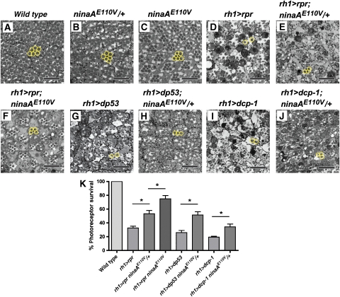
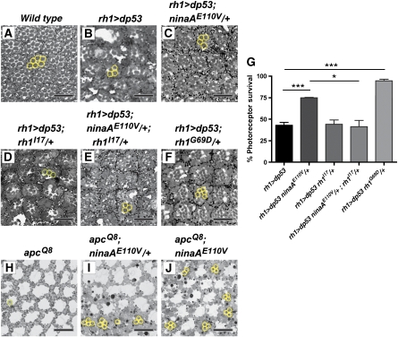
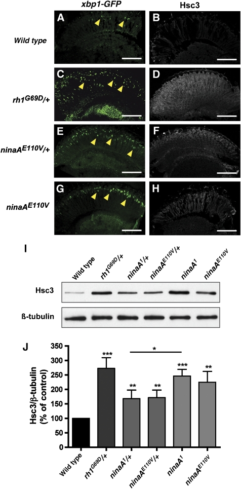
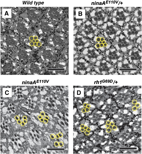
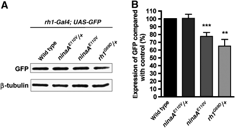
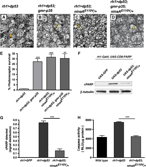
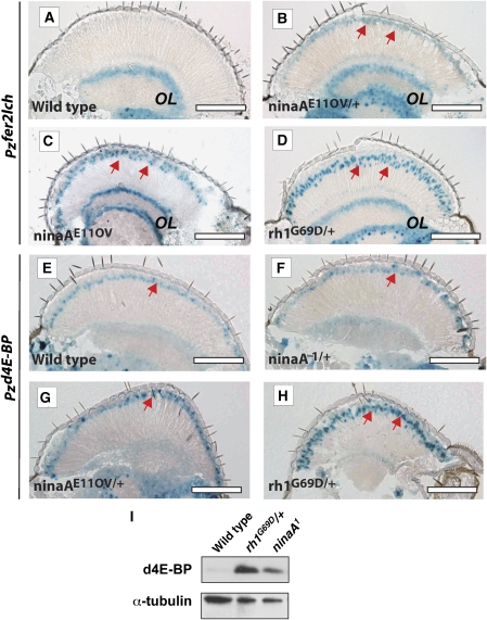
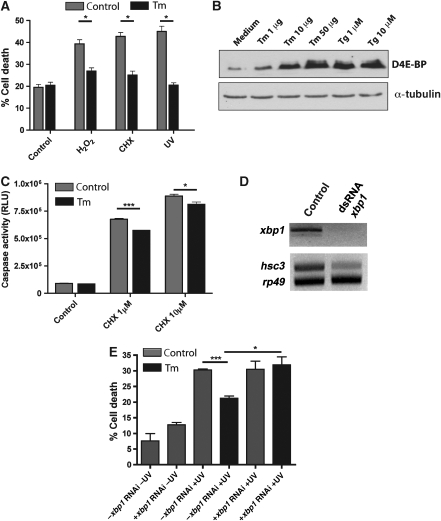
The unfolded protein response (UPR) is a specific cellular process that allows the cell to cope with the overload of unfolded/misfolded proteins in the endoplasmic reticulum (ER). ER stress is commonly associated with degenerative pathologies, but its role in disease progression is still a matter for debate. Here, we found that mutations in the ER-resident chaperone, neither inactivation nor afterpotential A (NinaA), lead to mild ER stress, protecting photoreceptor neurons from various death stimuli in adult Drosophila. In addition, Drosophila S2 cultured cells, when pre-exposed to mild ER stress, are protected from H(2)O(2), cycloheximide- or ultraviolet-induced cell death. We show that a specific ER-mediated signal promotes antioxidant defences and inhibits caspase-dependent cell death. We propose that an immediate consequence of the UPR not only limits the accumulation of misfolded proteins but also protects tissues from harmful exogenous stresses.


Figures

Figure 1
Figure 1
Loss of ninaA function protects PRCs from cell death. PRC integrity is visualized in outer PRCs submitted to the ectopic expression of rpr, dp53 and dcp-1, in a wild-type or ninaA mutant background. (AJ) PRC rhabdomeres are visualized in semi-thin tangential retinal sections. Scale bar, 10 μm. Outer PRC rhabdomeres of a representative ommatidium are circled in yellow. (A–C) PRC rhabdomeres are organized in a trapezoidal pattern in control retinas (5-day-old flies). (A) Wild type. (B) ninaAE110V/+. (C) ninaAE110V. (D–F) Loss of ninaA protects from rpr-induced cell death (1-day-old flies). (D) Ectopic expression of rpr in outer PRCs with rh1-Gal4;uas-rpr (rh1>rpr) induces a massive loss of PRC integrity. (E) Reduced PRC killing in rh1>rpr;ninaAE110V/+. (F) Reduced PRC killing in rh1>rpr;ninaAE110V. (G, H) Loss of ninaA protects from dp53-induced cell death (8-day-old flies). (G) Ectopic expression of dp53 in outer PRCs with rh1-Gal4;uas-dp53 (rh1>dp53) induces a massive PRC killing. (H) Reduced PRC killing in rh1>dp53/ninaAE110V/+. (I, J) Loss of ninaA protects from dcp-1-induced cell death (1-day-old flies). (I) Loss of PRCs in rh1-Gal4;;uas-dcp-1 (rh1>dcp-1). (J) Reduced killing in rh1-Gal4; ninaAE110V/+;uas-dcp-1. (K) Quantitative analysis of photoreceptor survival seen in (A, D–J). The results are expressed as mean percentage of wild-type control ±s.e.m. of three animals (*P<0.05 in Student's t-test).
Figure 2
Figure 2
The protective effect induced by the loss of ninaA is dependent on Rh1 levels. (AF) Semi-thin tangential retina sections of 2-day-old flies. Scale bar, 10 μm. Outer PRC rhabdomeres of representative ommatidium are surrounded by a yellow circle. (A) Wild type. (B) Ectopic expression of dp53 in outer PRCs (rh1>dp53) induces PRC killing. (C) PRC killing is reduced in rh1>dp53; ninaAE110V/+. (D) Reduced rh1 dosage does not protect from dp53-induced PRC killing (rh1>dp53; rh1I17/+). (E) Reduced rh1 dosage suppresses ninaAE110V/+ protective effect in PRCs submitted to dp53-induced killing (rh1>dp53; ninaAE110V/+; rh1I17/+). (F) A missense mutation in rh1 protects from dp53-induced PRC killing (rh1>dp53; rh1G69D/+). (G) Quantitative analysis of photoreceptor survival seen in (A–F). Results are expressed as mean percentage of survival ±s.e.m. of three animals (*P<0.05; ***P<0.001 in Student's t-test). (HJ) Loss of ninaA protects from apcQ8-induced cell death (1-day-old flies). Scale bar, 10 μm. (H) apcQ8 flies display no PRC. (I, J) Loss of ninaA reduces cell death induced by apcQ8 seen by the presence of rhabdomeres (yellow circles). (I) apcQ8; ninaAE110V/+. (J) apcQ8; ninaAE110V. Representative images of three animals.
Figure 3
Figure 3
xbp1 splicing and Hsc3 expression are increased in ninaA mutant retinas. Horizontal retina cryosections are stained with an antibody raised against GFP (A, C, E, G) or against Hsc3 (B, D, F, H) in 7-day-old flies. xbp1:GFP is expressed in PRCs under the control of rh1 promoter (rh1>xbp1:GFP). Scale bar, 100 μm. (A) Wild-type PRCs expressing xbp1:GFP show weak GFP staining. (C) rh1G69D/+, (E) ninaAE110V/+or (G) ninaAE110V retina expressing xbp1:GFP display an increased GFP staining in PRC nuclei (yellow arrowheads). (B) Wild-type retinas do not exhibit Hsc3 staining. (D) rh1G69D/+ retinas show a marked Hsc3 staining. (F) ninaAE110V/+ and (H) ninaAE110V retinas show weak Hsc3 staining. (I) Western blot analysis performed on fly eye tissue. ninaAE110V/+ or ninaA1/+ exhibit a clear Hsc3 increase compared with wild-type retina. Hsc3 protein level is further increased in ninaA1 compared with ninaA1/+. The Hsc3 level in rh1G69D/+ is increased to a level similar to that in ninaA amorphs. Representative blot of four independent experiments. (J) Quantification of Hsc3 immunoreactivity. The results are expressed as mean percentage of wild-type control ±s.e.m. of four independent experiments (*P<0.05; **P<0.01; ***P<0.001 in Student's t-test).
Figure 4
Figure 4
Mild and long-term accumulation of unfolded Rh1 protein does not promote age-related retinal degeneration. (AD) Semi-thin tangential retina sections in 60-day-old flies raised at 25°C in a 12:12 light–dark cycle. Outer PRC rhabdomeres of representative ommatidium are circled in yellow. Scale bar, 10 μm. (A) Wild type and (B) ninaAE110V/+ retinas exhibit a preserved retina structure with intact PRC rhabdomeres (100% ±0.0 PRC survival). (C) ninaAE110V (98.37% ±0.74 PRC survival) and (D) rh1G69D/+ (95.94% ±1.88 PRC survival) retinas show occasional signs of degeneration.
Figure 5
Figure 5
Effect of ER stress on the GAL4/UAS ectopic expression system. (A) Western blot analysis against GFP on head extracts expressing GFP under the control of the rh1-Gal4 driver in wild-type, ninaAE110V/+, ninaAE110V or rh1G69D/+ backgrounds. Representative blot from eight independent experiments. (B) Quantification of the amount of GFP detected in (A), normalized to the loading control and to the amount of GFP expressed in a wild-type background. There is no reduction of GFP protein levels in ninaAE110V/+ compared with wild type. ninaAE110V or rh1G69D/+ exhibit reproducible reductions in GFP levels compared with wild-type tissues. Reprobing with β-tubulin antibody serves as a loading control. The results are expressed as a mean percentage of wild-type control ±s.e.m. of eight independent experiments (**P<0.01; ***P<0.001 in Student's t-test).
Figure 6
Figure 6
Caspase activation is inhibited in the ninaA mutant retina. (AD) Semi-thin tangential retinal sections of 13-day-old flies expressing dp53 in outer photoreceptors cells. Outer PRC rhabdomeres of representative ommatidium are circled in yellow. Scale bar, 10 μm. (A) Ectopic expression of dp53 (rh1>dp53) induces outer PRC, but not inner PRC death that do not express dp53 (orange arrowhead). (B) dp53-induced PRC death is reduced by p35 concomitant expression (rh1>dp53; gmr-p35). (C) dp53-induced PRC death is reduced in ninaA mutants (rh1>dp53; ninaAE110V/+). (D) dp53-induced PRC death is reduced by p35 expression in the ninaA mutant (rh1>dp53; ninaAE110V/+; gmr-p35). (E) Quantitative analysis of photoreceptor survival seen in (A–D). The results are expressed as mean percentage of survival ±s.e.m. of three animals. (F) Caspase activity measured by the appearance of a cleaved PARP (cPARP) epitope by western blot using an anti-cPARP antibody. cPARP is present in tissue expressing dp53 in wild type but strongly reduced in ninaAE110V/+ background. Reprobing with β-tubulin antibody serves as loading control. Representative blot of three independent experiments. (G) Quantification of cPARP immunoreactivity. The results are expressed as mean percentage of total ±s.e.m. of three independent experiments. (H) Caspase activity measured by luminescence activity. The results are expressed as luminescence signal relative to the amount of protein±s.e.m. of three independent experiments (**P<0.01; ***P<0.001 in Student's t-test).
Figure 7
Figure 7
ER stress triggers upregulation of the fer2lch and d4E-BP genes. (AH) Horizontal retinal cryosections stained for nuclear β-galactosidase activity from flies carrying the transposable P-element line inserted in fer2lch (Pz[ry, lacZ]fer2lch035) (A–D) or d4E-BP (Pz[ry, lacZ ]thorl(2)06270) (E–H). Arrows point to nuclear β-galactosidase activity staining in PRC nuclei (B–H). (A) Wild-type retinas exhibit a fer2lch basal background staining in the optic lobe but no apparent staining in the retina. (B) ninaAE110V/+, (C) ninaAE110V and (D) rh1G69D/+ retinas exhibit increased β-galactosidase activity staining at the level of outer PRC nuclei. (E) Wild-type retina exhibit some d4E-BP staining in nuclei. d4E-BP expression is merely increased in ninaA1/+ (F) and ninaAE110V/+ (G) mutant retinas, but a marked increase is observed in rh1G69D/+ (H). Scale bar, 100 μm. (I) Western blot analysis of d4E-BP protein levels in rh1G69D/+ and ninaA1 compared with wild-type fly eye tissues. Representative blot of three independent experiments.
Figure 8
Figure 8
Mild ER stress protects S2 cells from oxidative and apoptotic insults. (A) Percentage of cell death was evaluated by trypan blue uptake in untreated (control, gray columns) and pre-treated S2 cells with 1 μg/ml of Tm (black columns), which were then submitted to H2O2 (500 nM), CHX (1 μM) or UVC exposure (300 mJ/cm2). The results are expressed as mean percentage of cell death ±s.e.m. of three independent experiments. (B) Tm or Tg treatment increased d4E-BP expression in a dose-dependent manner. d4E-BP expression is visualized by western blot analysis on S2 cells treated with Tm or Tg for 6 h. (C) Tm pre-treatment reduces caspase activity induced by CHX treatment. Cells were either untreated (control, gray columns) or pre-treated with 1 μg/ml of Tm (black columns) then submitted to CHX (1 and 10 μM). The results are expressed as RLU (relative luminescent unit) ±s.e.m. of one experiment (triplicates) representative of three independent experiments. (D) xbp1 knock down by dsRNA strongly reduced the amount of xbp1 and hsc3 transcripts. The expression of the ribosomal rp49 gene was used as internal control for RNA extraction and RT–PCR efficiency. Its expression is constant in control and treated cells. (E) Knock down of xbp1 reverts the protective effect of Tm. The results are expressed as mean percentage of cell death ±s.e.m. of three independent experiments. (in A, B, C and E, *P<0.05; **P<0.01; ***P<0.001 in Student's t-test).

Similar articles

Cited by

References

    1. Ahmed Y, Hayashi S, Levine A, Wieschaus E (1998) Regulation of armadillo by a Drosophila APC inhibits neuronal apoptosis during retinal development. Cell 93: 1171–1182 - PubMed
    1. Baker EK, Colley NJ, Zuker CS (1994) The cyclophilin homolog NinaA functions as a chaperone, forming a stable complex in vivo with its protein target rhodopsin. EMBO J 13: 4886–4895 - PMC - PubMed
    1. Boyce M, Bryant KF, Jousse C, Long K, Harding HP, Scheuner D, Kaufman RJ, Ma D, Coen DM, Ron D, Yuan J (2005) A selective inhibitor of eIF2alpha dephosphorylation protects cells from ER stress. Science 307: 935–939 - PubMed
    1. Brodsky MH, Nordstrom W, Tsang G, Kwan E, Rubin GM, Abrams JM (2000) Drosophila p53 binds a damage response element at the reaper locus. Cell 101: 103–113 - PubMed
    1. Calfon M, Zeng H, Urano F, Till JH, Hubbard SR, Harding HP, Clark SG, Ron D (2002) IRE1 couples endoplasmic reticulum load to secretory capacity by processing the XBP-1 mRNA. Nature 415: 92–96 - PubMed

Publication types

MeSH terms