. 2009;4(4):e5160.
doi: 10.1371/journal.pone.0005160. Epub 2009 Apr 2.

Targeting and function of the mitochondrial fission factor GDAP1 are dependent on its tail-anchor

Affiliations

Targeting and function of the mitochondrial fission factor GDAP1 are dependent on its tail-anchor

Konstanze M Wagner et al. PLoS One. 2009.

Abstract

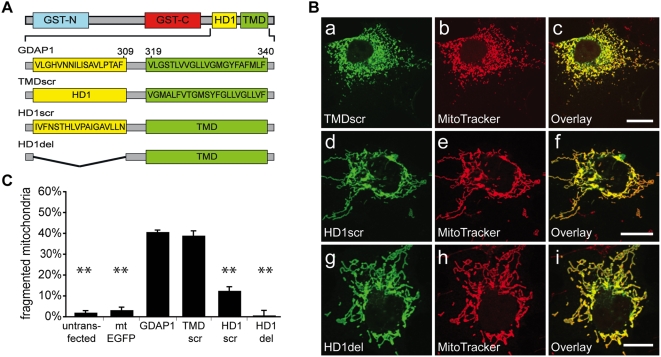
Proteins controlling mitochondrial dynamics are often targeted to and anchored into the mitochondrial outer membrane (MOM) by their carboxyl-terminal tail-anchor domain (TA). However, it is not known whether the TA modulates protein function. GDAP1 is a mitochondrial fission factor with two neighboring hydrophobic domains each flanked by basic amino acids (aa). Here we define GDAP1 as TA MOM protein. GDAP1 carries a single transmembrane domain (TMD) that is, together with the adjacent basic aa, critical for MOM targeting. The flanking N-terminal region containing the other hydrophobic domain is located in the cytoplasm. TMD sequence, length, and high hydrophobicity do not influence GDAP1 fission function if MOM targeting is maintained. The basic aa bordering the TMD in the cytoplasm, however, are required for both targeting of GDAP1 as part of the TA and GDAP1-mediated fission. Thus, this GDAP1 region contains critical overlapping motifs defining intracellular targeting by the TA concomitant with functional aspects.


Conflict of interest statement

Competing Interests: The authors have declared that no competing interests exist.

Figures

Figure 1
Figure 1. GDAP1 membrane topology.
(A) Graphic of experimental strategy. (B) Western blot of mitochondria-enriched fractions derived from GDAP1-FLAG-transfected HeLa cells treated with proteinase K for 30 minutes at 4°C with increasing digitonin concentrations. Without digitonin, the cytosolic N-terminal part of GDAP1-FLAG is degraded (arrow) and only the C-terminal 5 kDa fragment is detected (arrowhead). After membrane permeabilization, the C-terminal fragment is gradually degraded confirming its location in the IMS. The IMS protein Opa1 served as control. (C) Confocal immunofluorescence microscopy of transfected COS-7 cells expressing GDAP1-FLAG (a–d). Co-stainings: MitoTracker (mitochondria). Bars, 10 µm.
Figure 2
Figure 2. Integration assay of in vitro-synthesized TA-proteins.
(A) The post-nuclear supernatant of HeLa cells was incubated with the in vitro-translates of GDAP1, Fis1, Mfn2, Luciferase, EGFP-HD1-TMD or EGFP-TMD. Mitochondria were enriched in a differential centrifugation approach (S1, P1, S2, P2). GDAP1, Fis1, EGFP-HD1-TMD and EGFP-TMD showed co-sedimentation with the mitochondrial marker porin. Mfn2 and Luciferase remained in the supernatant. (B) The post-nuclear supernatant of HeLa cells was incubated with the in vitro-translated GDAP1, Fis1, EGFP-HD1-TMD and EGFP-TMD and the mitochondrial pellet was resuspended in buffer (control), in 1 M NaCl, 0.1 M carbonate (pH 11), or in buffer with 0.1% TritonX-100, and centrifuged to separate the soluble protein supernatants (S) from membranous pellets (P). All in vitro-translated proteins and the integral membrane protein porin could be sedimented and became soluble only upon treatment with detergent. Unlike the integral membrane proteins cytochrome C can only be sedimented under control conditions. (C) Immunoprecipitation of in vitro translated GDAP1-FLAG with an anti-FLAG antibody without and after membrane integration and with or without proteinase K (50 mg/ml) digest. The upper arrow points to the undigested immuno-precipitated full length GDAP1-FLAG. The lower arrows indicates the 5 kD fragment still detected after membrane integration and protease digest.
Figure 3
Figure 3. TA-associated critical basic residues for GDAP1 localization and function.
(A) Schema of GDAP1 and aa sequence of C-terminal part of wt GDAP1 (GDAP1) and mutants (bold letters: altered aa sequence). (B) Confocal immunofluorescence microscopy of transfected COS-7 cells expressing mtEGFP as control (a–d) wt GDAP1 (e–h) and mutant proteins (i–x). Co-stainings: MitoTracker (mitochondria), PDI (ER). Bars, 10 µm. (C) Quantification of GDAP1 and mutants colocalized with MitoTracker. (D) Quantification of mitochondrial morphology.
Figure 4
Figure 4. Role of TMD and HD1 amino acid sequences in GDAP1-mediated fission.
(A) Schematic representation of the aa sequence of the TMD and HD1 of wt GDAP1 (GDAP1) and mutants TMDscr, HD1scr and HD1del. (B) Distinct mitochondrial localization of mutant proteins in transfected COS-7 cells (a–i). (C) Quantification of mitochondrial morphology. Fission activity was lost by HD1 scrambling or deletion. Bars, 10 µm.
Figure 5
Figure 5. Function of TA in GDAP1 fission activity.
(A) Construction of chimera GDAP1-VAMP1B. (B) COS-7 cells transfected with GDAP1-VAMP1B display mitochondrial localization and fragmented mitochondria comparable to full-length GDAP1 (a–c). (C) Quantification reveals no difference in fission activity between GDAP1 and GDAP1-Vamp1B. Bars, 10 µm.
Figure 6
Figure 6. Influence of TMD hydrophobicity on GDAP1 fission activity.
(A) COS-7 cells expressing either GDAP1-OMb5 (a–c) or GDAP1hy (d–f) were co-stained with MitoTracker. Both proteins show mitochondrial localization, with some mislocalization of GDAP1-OMb5 to the ER (for quantification, see Suppl. Fig. A3). (B) Quantification of mitochondrial morphology. Bars, 10 µm.
Figure 7
Figure 7. Model of possible GDAP1 membrane topologies.
Experimentally confirmed is a single transmembrane span with the C-terminus in the intermembrane space and the N-terminus in the cytosol. Within this topology two different arrangements of the HD1 are represented. Crossed-out: Experimentally disproved topology. MOM, mitochondrial outer membrane.

References

    1. Chan DC. Mitochondria: dynamic organelles in disease, aging, and development. Cell. 2006;125:1241–1252. - PubMed
    1. Niemann A, Berger P, Suter U. Pathomechanisms of mutant proteins in Charcot-Marie-Tooth disease. Neuromolecular Med. 2006;8:217–242. - PubMed
    1. Waterham HR, Koster J, van Roermund CW, Mooyer PA, Wanders RJ, et al. A lethal defect of mitochondrial and peroxisomal fission. N Engl J Med. 2007;356:1736–1741. - PubMed
    1. Rojo M, Legros F, Chateau D, Lombes A. Membrane topology and mitochondrial targeting of mitofusins, ubiquitous mammalian homologs of the transmembrane GTPase Fzo. J Cell Sci. 2002;115:1663–1674. - PubMed
    1. Borgese N, Colombo S, Pedrazzini E. The tale of tail-anchored proteins: coming from the cytosol and looking for a membrane. J Cell Biol. 2003;161:1013–1019. - PMC - PubMed

Publication types