. 2009;4(4):e5045.
doi: 10.1371/journal.pone.0005045. Epub 2009 Apr 2.

High-fat diet induces apoptosis of hypothalamic neurons

Affiliations

High-fat diet induces apoptosis of hypothalamic neurons

Juliana C Moraes et al. PLoS One. 2009.

Expression of concern in

Abstract

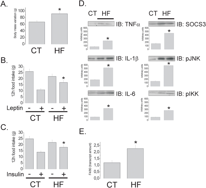
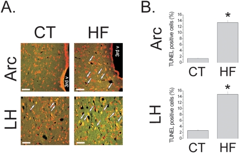
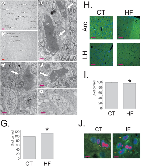
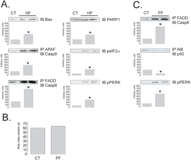
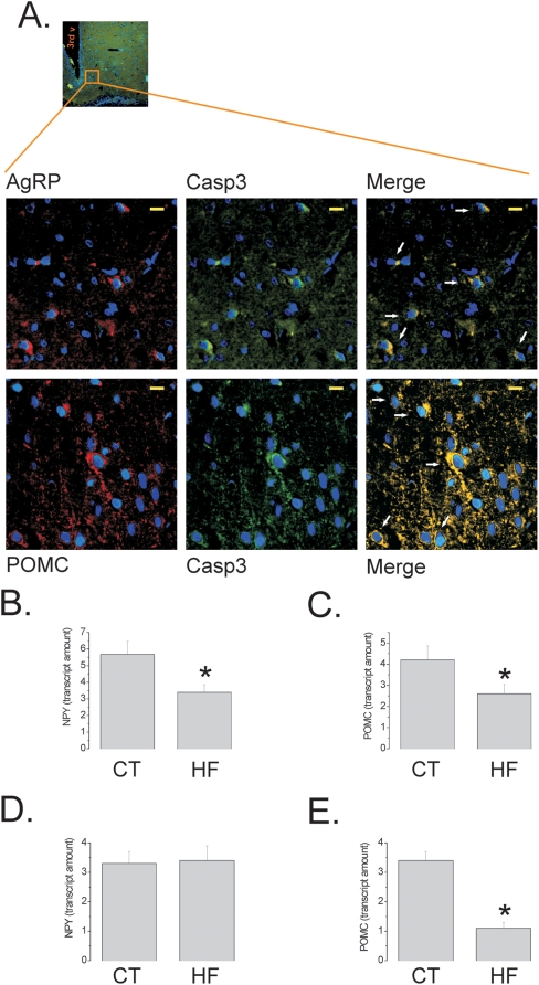
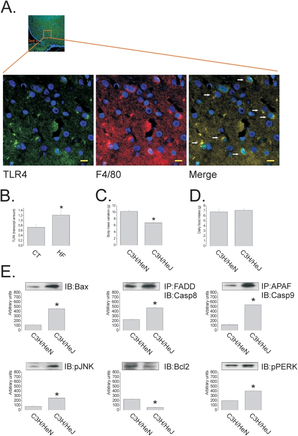
Consumption of dietary fats is amongst the most important environmental factors leading to obesity. In rodents, the consumption of fat-rich diets blunts leptin and insulin anorexigenic signaling in the hypothalamus by a mechanism dependent on the in situ activation of inflammation. Since inflammatory signal transduction can lead to the activation of apoptotic signaling pathways, we evaluated the effect of high-fat feeding on the induction of apoptosis of hypothalamic cells. Here, we show that consumption of dietary fats induce apoptosis of neurons and a reduction of synaptic inputs in the arcuate nucleus and lateral hypothalamus. This effect is dependent upon diet composition, and not on caloric intake, since pair-feeding is not sufficient to reduce the expression of apoptotic markers. The presence of an intact TLR4 receptor, protects cells from further apoptotic signals. In diet-induced inflammation of the hypothalamus, TLR4 exerts a dual function, on one side activating pro-inflammatory pathways that play a central role in the development of resistance to leptin and insulin, and on the other side restraining further damage by controlling the apoptotic activity.


Conflict of interest statement

Competing Interests: The authors have declared that no competing interests exist.

Figures

Figure 1
Figure 1. Leptin/insulin resistance and inflammatory markers in the hypothalamus of rats fed on high-fat diet.
(A) Body mass variation (g) of Wistar rats fed on control (CT) or high-fat (HF) diets for 8 w. (B–C) Twelve hours spontaneous food intake (g) of Wistar rats fed on CT or HF diets for 8 w and treated icv with a single dose (2.0 µl) of saline (−), leptin (+, in B) or insulin (+, in C). (D) Immunoblots (IB) of hypothalamic protein extracts obtained from Wistar rats fed on CT or HF diets. (E) Real-time PCR analysis of F4/80 transcript amount in samples obtained from the hypothalami of Wistar rats fed on CT or HF diets. In all experiments n = 5. In A, D and E, *p<0.05 vs. CT, values are means±SEM; in B and C, *p<0.05 vs. CT+, values are means±SEM.
Figure 2
Figure 2. TUNEL assay depicts apoptosis in the hypothalamus of rats fed on high-fat diet.
(A) Representative microphotographs of DNA fragmentation detection by TUNEL (stained in yellow) in samples from arcuate (Arc) and lateral hypothalamic (LH) nuclei, from Wistar rats fed on control (CT) or high-fat (HF) diets; the arrows indicate TUNEL positive cells. (B–C) TUNEL positive cells in Arc (B) and LH (C) are expressed as % of total cells per field. In all experiments n = 5. In A, magnification, ×200 (scale bar, 20 µm). In B and C, *p<0.05 vs. CT, values are means±SEM.
Figure 3
Figure 3. Apoptotic neurons in the hypothalamus of rats fed on high-fat diet.
(A–F) Transmission electron microscopy images of typical apoptotic neurons (A–E) and a normal neuron (F) in the arcuate nucleus of Wistar rats fed on high-fat (A, C–E) and control (B, F) diets, respectively. The arrows indicate typical apoptotic neurons. (G) Apoptotic neurons were counted in low magnification fields of transmission electron microscopy analysis from the arcuate nucleus of Wistar rats fed on high-fat (HF) and control (CT) diets, the results are presented as % of CT. (H) Representative synaptophysin immunofluorescence staining of samples from arcuate (Arc) and lateral hypothalamic (LH) nuclei, from Wistar rats fed on CT or HF diets. (I) Synaptophysin positive nerve terminals were field counted and the results are presented as % of CT. (J) Representative NeuN (rhodamine) and Bax (fluorescein) double immunoflorescence staining of samples from hypothalamus of Wistar rats fed on CT and HF diets; orange arrows depict neurons without Bax expression, yellow arrows depict double positive NeuN/Bax stained neurons. A–F are representative of n = 3; magnification, ×100 (scale bar, 20 µm), A–B; and ×20,000 (scale bar, 0.2 µm), C–F. G, field counting was performed in five distinct fields from n = 3; *p<0.05 vs. CT. H and J are representative of n = 5; magnification, ×100 (scale bar, 40 µm), H; and ×400 (scale bar, 10 µm), J; nuclei are stained in blue by DAPI. I, nerve terminals were counted in120 distinct fields (×200 magnification) from n = 5; *p<0.05 vs. CT.
Figure 4
Figure 4. Apoptotic and endoplasmic reticulum stress markers in the hypothalamus of rats fed on high-fat diet.
(A and C) Immunoblots (IB) of hypothalamic protein extracts obtained from rats fed control (CT), high-fat (HF) (A) or HF diet in pair-feeding (PF) (C); in some cases samples were submitted to immunoprecipitation (IP) prior to IB. (B) Body mass variation (g) of Wistar rats fed on CT or HF diet in pair-feeding (PF) for 8 w. In all experiments, n = 5; *p<0.05 vs. CT.
Figure 5
Figure 5. Differences in neuronal subpopulation apoptosis in diet-induced obesity.
(A) Representative AgRP (rhodamine) and Caspase-3 (Casp3, fluorescein) (upper panels) or POMC (rhodamine) and Caspase-3 (Casp3, fluorescein) (lower panels) double immunoflorescence staining of samples from hypothalamus of Wistar rats fed on high-fat diet; arrows in merge depict double positive neurons; inset depicts approximate site in the arcuate nucleus that was evaluated in detail. (B–E) Real-time PCR analysis of NPY (B and D) and POMC (C and E) transcript amounts in samples obtained from the hypothalami of Wistar rats (B–C) and Swiss mice (D–E) fed on control (CT) or high-fat (HF) diets. In all experiments n = 5. In A, inset magnification, ×20 and captions magnification, ×400 (scale bar, 10 µm), nuclei are stained in blue by DAPI; 3rd v, third ventricle. In B–E, *p<0.05 vs. CT.
Figure 6
Figure 6. TLR4 protects against diet-induced apoptosis of hypothalamic neurons.
(A) Representative TLR4 (rhodamine) and F4/80 (fluorescein) double immunoflorescence staining of samples from hypothalamus of Wistar rats; arrows in merge depict double positive cells; inset depicts approximate site in the arcuate nucleus that was evaluated in detail. (B) Real-time PCR analysis of TLR4 transcript amount in samples obtained from the hypothalami of Wistar rats fed on control (CT) or high-fat (HF) diets. (C) Body mass variation (g) of C3H/HeN and C3H/HeJ mice fed on HF diet for 8 w. (D) Mean daily food intake (g) of C3H/HeN and C3H/HeJ mice fed on HF diet. (E) Immunoblots (IB) of hypothalamic protein extracts obtained from C3H/HeN and C3H/HeJ mice fed on HF diet; in some cases samples were submitted to immunoprecipitation (IP) prior to IB. In all experiments n = 5. In A, inset magnification, ×20 and captions magnification, ×400 (scale bar, 10 µm), nuclei are stained in blue by DAPI; 3rd v, third ventricle. B, *p<0.05 vs. CT; in C and E, *p<0.05 vs. C3H/HeN.

References

    1. Flier JS. Obesity wars: molecular progress confronts an expanding epidemic. Cell. 2004;116:337–350. - PubMed
    1. Milanski M, Degasperi G, Coope A, Morari J, Denis R, et al. Saturated fatty acids produce an inflammatory response predominantly through the activation of TLR4 signaling in hypothalamus: implications for the pathogenesis of obesity. J Neurosci. 2009;29:359–370. - PMC - PubMed
    1. De Souza CT, Araujo EP, Bordin S, Ashimine R, Zollner RL, et al. Consumption of a fat-rich diet activates a proinflammatory response and induces insulin resistance in the hypothalamus. Endocrinology. 2005;146:4192–4199. - PubMed
    1. Munzberg H, Flier JS, Bjorbaek C. Region-specific leptin resistance within the hypothalamus of diet-induced obese mice. Endocrinology. 2004;145:4880–4889. - PubMed
    1. Howard JK, Cave BJ, Oksanen LJ, Tzameli I, Bjorbaek C, et al. Enhanced leptin sensitivity and attenuation of diet-induced obesity in mice with haploinsufficiency of Socs3. Nat Med. 2004;10:734–738. - PubMed

Publication types