

One-stage surgical management for children with spinal tuberculosis by anterior decompression and posterior instrumentation
- PMID: 19340427
- PMCID: PMC2899114
- DOI: 10.1007/s00264-009-0758-5
One-stage surgical management for children with spinal tuberculosis by anterior decompression and posterior instrumentation
Abstract
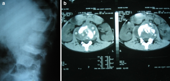
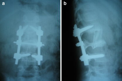
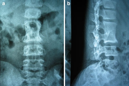
The goal of this study was to assess the efficacy of one-stage surgical management for children with spinal tuberculosis by anterior decompression, bone grafting, posterior instrumentation, and fusion. Between January 2002 and December 2006, 15 cases with spinal tuberculosis were treated with one-stage posterior internal fixation and anterior debridement. All cases were followed-up for an average of 30.3 months (range 12-48 months). The average neurological recovery in the patients was 0.93 grades on the scale of Frankel et al. (Paraplegia 7:179-192, 1969). The average preoperative kyphosis was 36 degrees (range 19-59 degrees ), and the average postoperative kyphosis was 23 degrees (range 15-38 degrees ) at final follow-up. At final follow-up, minimal progression of kyphosis was seen, with an average kyphosis of 27 degrees (range 16-40 degrees ). An average loss of correction of 4 degrees was seen at final follow-up. One-stage surgical management for children with spinal tuberculosis by anterior decompression, bone grafting, posterior instrumentation, and fusion was feasible and effective.
Figures
Comment in
-
Comment on Huang et al.: One-stage surgical management for children with spinal tuberculosis by anterior decompression and posterior instrumentation.Int Orthop. 2010 Jun;34(5):769-70; author relply 771. doi: 10.1007/s00264-010-0975-y. Epub 2010 Feb 20. Int Orthop. 2010. PMID: 20174795 Free PMC article. No abstract available.
References
-
- Boachie-Adjei O, Squillante RG. Tuberculosis of the spine. Orthop Clin North Am. 1996;27:95–103. - PubMed
-
- Frankel HL, Hancock DO, Hyslop G, et al. The value of postural reduction in the initial management of closed injuries of the spine with paraplegia and tetraplegia. Paraplegia. 1969;7:179–192. - PubMed
MeSH terms
Substances
LinkOut - more resources
Full Text Sources
