

Fundus autofluorescence in cone dystrophy
- PMID: 19340470
- PMCID: PMC2752495
- DOI: 10.1007/s10633-009-9172-y
Fundus autofluorescence in cone dystrophy
Abstract
Purpose: To describe fundus autofluorescence (FAF) finding in a case of cone dystrophy.
Methods: Interventional case report.
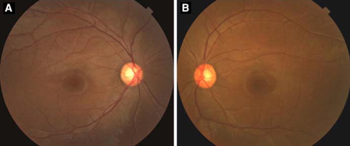
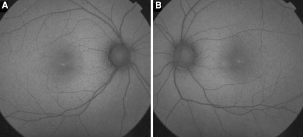
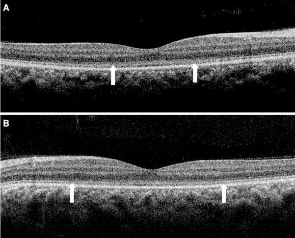
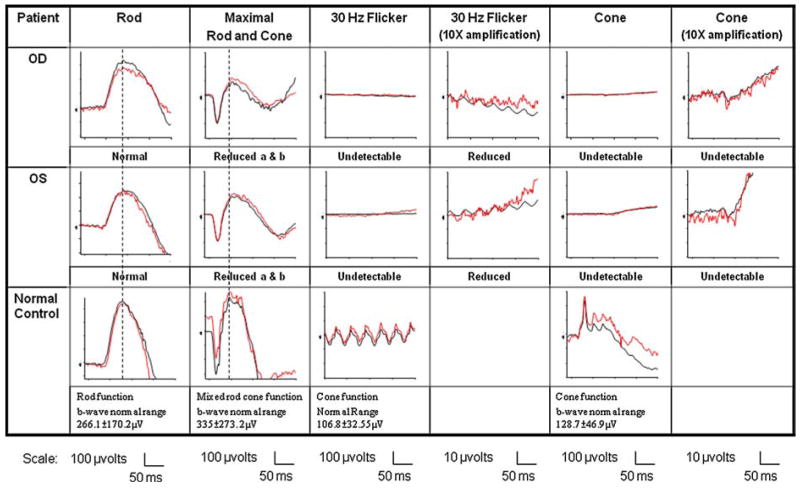
Results: A 23-year-old woman presented with increasing photophobia and decreasing vision in both eyes for 2 years. Fundus examination showed several drusen-like dots. FAF revealed hyper-autofluorescence in the foveola. Electroretinogram (ERG) demonstrated a pure "cone" dystrophy.
Conclusion: Hyper-autofluorescence in the foveola is a non-specific manifestation of photoreceptor-retinal pigment epithelium dysfunction. ERG studies are essential for accurate diagnosis.
Figures
References
-
- Kurz-Levin MM, Halfyard AS, Bunce C, Bird AC, Holder GE. Clinical variations in assessment of bull’s-eye maculopathy. Arch Ophthalmol. 2002;120:567–575. - PubMed
-
- Holz FG, Spaide RF, Bird AC. Atlas of fundus autofluorescence imaging. 1. Springer; New York: 2007. pp. 199–205.
-
- Krill AE, Deutman AF. Dominant macular degenerations. The cone dystrophies. Am J Ophthalmol. 1972;73:352–369. - PubMed
-
- Deutman AF. Benign concentric annular macular dystrophy. Am J Ophthalmol. 1974;78:384–396. - PubMed
Publication types
MeSH terms
Grants and funding
LinkOut - more resources
Full Text Sources
Medical
