. 2009 Jul;30(5):710-5.
doi: 10.1007/s00246-009-9408-3. Epub 2009 Apr 2.

Cell-cycle-based strategies to drive myocardial repair

Affiliations

Cell-cycle-based strategies to drive myocardial repair

Wuqiang Zhu et al. Pediatr Cardiol. 2009 Jul.

Abstract

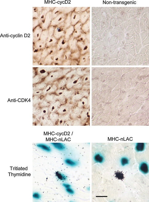
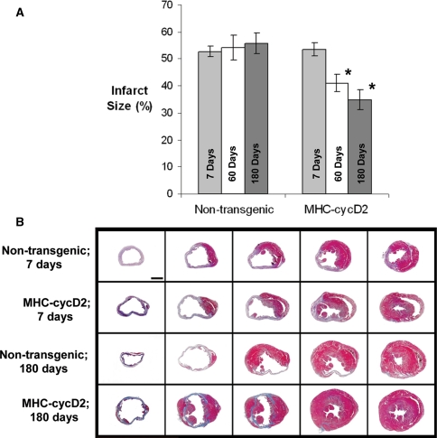
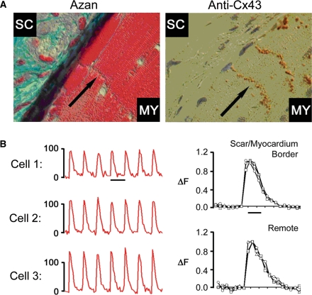
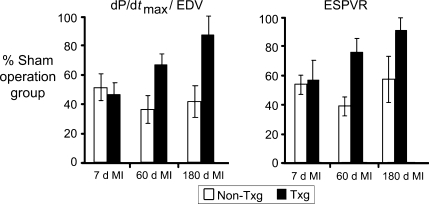
Cardiomyocytes exhibit robust proliferative activity during development. After birth, cardiomyocyte proliferation is markedly reduced. Consequently, regenerative growth in the postnatal heart via cardiomyocyte proliferation (and, by inference, proliferation of stem-cell-derived cardiomyocytes) is limited and often insufficient to affect repair following injury. Here, we review studies wherein cardiomyocyte cell cycle proliferation was induced via targeted expression of cyclin D2 in postnatal hearts. Cyclin D2 expression resulted in a greater than 500-fold increase in cell cycle activity in transgenic mice as compared to their nontransgenic siblings. Induced cell cycle activity resulted in infarct regression and concomitant improvement in cardiac hemodynamics following coronary artery occlusion. These studies support the notion that cell-cycle-based strategies can be exploited to drive myocardial repair following injury.


Figures

Fig. 1
Fig. 1
Transgene expression and cardiomyocyte DNA synthesis in postnatal MHC-cycD2 hearts. Heart sections from MHC-cycD2 mice and their nontransgenic littermates were subjected to anti-cyclin D2 (upper panels) and anti-CDK4 (middle panels) immune histology (horseradish peroxidase-conjugated secondary antibody, dark brown signal from diaminobenzidine reaction). Cyclin D2 expression results in a concomitant induction of the endogenous CDK4 gene product in transgenic hearts. Heart sections from MHC-cycD2/MHC-nLAC double transgenic mice and their MHC-nLAC single transgenic littermates were processed for cardiomyocyte DNA synthesis assay (mice received an injection of tritiated thymidine prior to sacrifice). The presence of silver grains over blue nuclei is indicative of cardiomyocyte DNA synthesis and is readily seen in sections from mice carrying the MHC-cycD2 transgene
Fig. 2
Fig. 2
Expression of cyclin D2 results in infarct regression. a Infarct size in nontransgenic and MHC-cycD2 transgenic mice at 7, 6, and 180 days postinjury. Asterisks indicate statistical significance between MHC-cycD2 hearts and. noninfarcted hearts at the indicated time point. b Representative sections from infarcted MHC-cycD2 transgenic hearts and their nontransgenic siblings at 7 and 180 days postinjury. Sections were sampled at 1-mm intervals from the apex to the base and were stained with Azan
Fig. 3
Fig. 3
Regenerated myocardium in MHC-cycD2 hearts is functionally integrated. Samples are from MHC-cycD2 hearts at 180 days postinfarction. a Apically located regenerated cardiomyocytes are well developed (left panel, Azan stain) and are interconnected via well-developed junctional complexes containing anti-connexin43 immune reactivity (right panel, horseradish peroxidase-conjugated secondary antibody, dark brown signal from diaminobenzidine reaction). b Intracellular calcium transients (as evidenced via changes in the fluorescence of the calcium-sensing dye rhod2) recorded from three apically located cardiomyocytes in the regenerated myocardium (left panels). Intracellular calcium transients in cardiomyocytes located in the regenerated myocardium were in synchrony with and indistinguishable from those in the remote myocardium (right panels)
Fig. 4
Fig. 4
Left ventricular performance was assessed by pressure–volume measurements. The left panel shows the left ventricular peak positive developed pressure/end diastolic volume (dP/dtmax/EDV) relationship for nontransgenic (open bars) and MHC-cycD2 mice (closed bars) at 7, 60, and 180 days after myocardial infarction. The values shown were normalized to those obtained from sham-operated mice from the same time points. The right panel shows the end systolic pressure–volume relationship (EDPVR)

Similar articles

Cited by

References

    1. Bergmann O, Bhardwaj RD, Bernard S, Zdunek S, Barnabé-Heider F, Walsh S, Zupicich J, Kanar Alkass, Buchholz BA, Druid H, Jovinge S, Frisen J (2008) Turnover of human cardiomyocytes. Circulation 118(Suppl):Abstract 3526
    1. {'text': '', 'ref_index': 1, 'ids': [{'type': 'DOI', 'value': '10.1074/jbc.M404975200', 'is_inner': False, 'url': 'https://doi.org/10.1074/jbc.m404975200'}, {'type': 'PubMed', 'value': '15159393', 'is_inner': True, 'url': 'https://pubmed.ncbi.nlm.nih.gov/15159393/'}]}
    2. Chaudhry HW, Dashoush NH, Tang H, Zhang L, Wang X, Wu EX, Wolgemuth DJ (2004) Cyclin A2 mediates cardiomyocyte mitosis in the postmitotic myocardium. J Biol Chem 279:35858–35866 - PubMed
    1. {'text': '', 'ref_index': 1, 'ids': [{'type': 'DOI', 'value': '10.1016/0092-8674(93)90137-F', 'is_inner': False, 'url': 'https://doi.org/10.1016/0092-8674(93)90137-f'}, {'type': 'PubMed', 'value': '8490963', 'is_inner': True, 'url': 'https://pubmed.ncbi.nlm.nih.gov/8490963/'}]}
    2. Dowdy SF, Hinds PW, Louie K, Reed SI, Arnold A, Weinberg RA (1993) Physical interaction of the retinoblastoma protein with human D cyclins. Cell 73:499–511 - PubMed
    1. {'text': '', 'ref_index': 1, 'ids': [{'type': 'DOI', 'value': '10.1101/gad.1306705', 'is_inner': False, 'url': 'https://doi.org/10.1101/gad.1306705'}, {'type': 'PMC', 'value': 'PMC1132004', 'is_inner': False, 'url': 'https://pmc.ncbi.nlm.nih.gov/articles/PMC1132004/'}, {'type': 'PubMed', 'value': '15870258', 'is_inner': True, 'url': 'https://pubmed.ncbi.nlm.nih.gov/15870258/'}]}
    2. Engel FB, Schebesta M, Duong MT, Lu G, Ren S, Madwed JB, Jiang H, Wang Y, Keating MT (2005) p38 MAP kinase inhibition enables proliferation of adult mammalian cardiomyocytes. Genes Dev 19:1175–1187 - PMC - PubMed
    1. {'text': '', 'ref_index': 1, 'ids': [{'type': 'DOI', 'value': '10.1073/pnas.0607382103', 'is_inner': False, 'url': 'https://doi.org/10.1073/pnas.0607382103'}, {'type': 'PMC', 'value': 'PMC1622860', 'is_inner': False, 'url': 'https://pmc.ncbi.nlm.nih.gov/articles/PMC1622860/'}, {'type': 'PubMed', 'value': '17032753', 'is_inner': True, 'url': 'https://pubmed.ncbi.nlm.nih.gov/17032753/'}]}
    2. Engel FB, Hsieh PC, Lee RT, Keating MT (2006) FGF1/p38 MAP kinase inhibitor therapy induces cardiomyocyte mitosis, reduces scarring, and rescues function after myocardial infarction. Proc Natl Acad Sci USA 103:15546–15551 - PMC - PubMed