. 2009 Jul;59(4):253-62.
doi: 10.1007/s12576-009-0030-1. Epub 2009 Mar 4.

The opposite roles of nNOS in cardiac ischemia-reperfusion-induced injury and in ischemia preconditioning-induced cardioprotection in mice

Affiliations

The opposite roles of nNOS in cardiac ischemia-reperfusion-induced injury and in ischemia preconditioning-induced cardioprotection in mice

Xiao-Mei Lu et al. J Physiol Sci. 2009 Jul.

Abstract

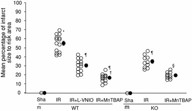
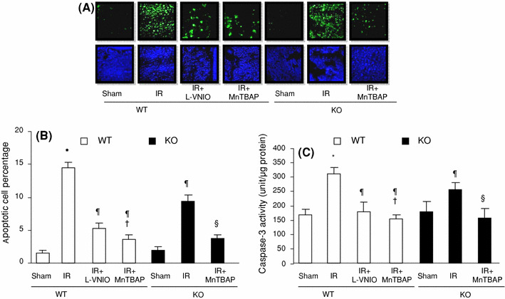
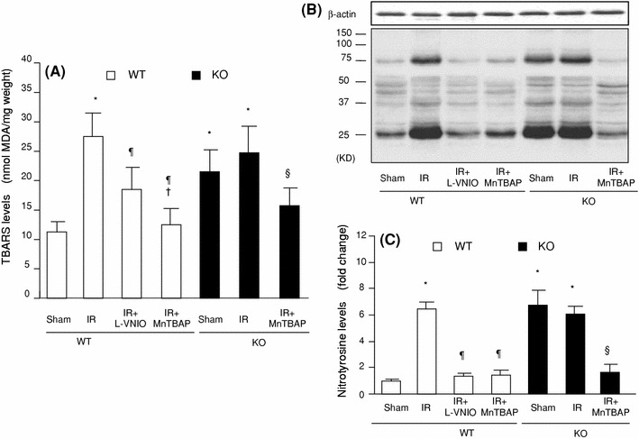
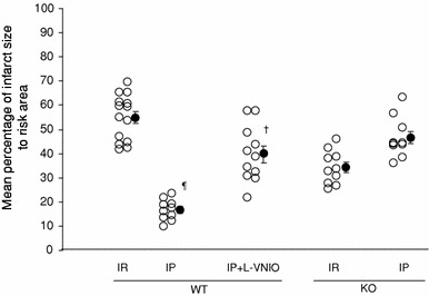
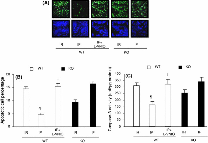
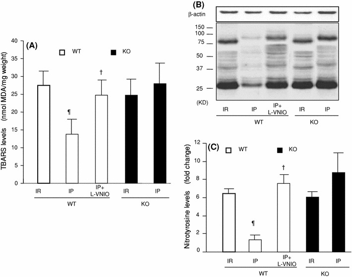
The role of neuronal nitric oxide synthase (nNOS) in cardiac ischemia-reperfusion (IR) and ischemia preconditioning (IP) is still controversial. Here, we focused on the possible roles of nNOS in cardiac IR and IP. Wild type C57BL/6 (WT) mice were subjected to coronary artery occlusion for 30 min followed by 24-h reperfusion (IR). Cardiac injury (infarct size and apoptotic cell number) was increased, associated with elevation of oxidative stress (lipid peroxidation) and nitrative stress (nitrotyrosine formation). A potent nNOS inhibitor, L-VNIO, and a superoxide dismutase mimetic and peroxynitrite scavenger, MnTBAP, significantly reduced IR-induced increases of oxidative/nitrative stress and cardiac injury. IR-induced cardiac injury in nNOS(-/-) (KO) mice was significantly lower than that in WT mice. MnTBAP markedly reduced IR-induced cardiac injury by suppression of oxidative/nitrative stress in KO mice. Cardiac IP was performed by three cycles of 5-min IR before 30-min ischemia followed by 24-h reperfusion. IP attenuated IR-induced cardiac injury in WT mice associated with reductions of oxidative/nitrative stress. IP-induced reduction of cardiac injury and oxidative/nitrative stress were eliminated by pretreatment with L-VNIO. In contrast with WT mice, IP had no protective effects in nNOS KO mice. In conclusion, nNOS played a dual role during cardiac IR and IP; nNOS exacerbated IR-induced injury by increasing oxidative/nitrative stress and contributed to IP-induced protection by inhibition of oxidative/nitrative stress.


Figures

Fig. 1
Fig. 1
Effects of L-VNIO and MnTBAP on IR-induced cardiac infarct size. Cardiac infarct size was measured as described in “Methods”. Results are expressed as mean percentage of infarct size to risk area. WT denotes wild type and KO denotes nNOS−/−. Open circles denote individual mice whereas solid circles denote mean ± SEM. *< 0.05 compared with the WT Sham group, ¶< 0.05 compared with the WT IR group, †< 0.05 compared with the WT IR + LVNIO group, §< 0.05 compared with the KO IR group
Fig. 2
Fig. 2
Effects of L-VNIO and MnTBAP on IR-induced cardiac apoptosis. a Representative TUNEL-positive staining for each group, as described in “Methods”. Green spots indicate positive apoptosis nuclei and blue indicates individual nuclei. b Percentage of positive apoptosis nuclei and total nuclei expressed as mean ± SEM. c Caspase-3 activities were measured as described in “Methods”, and are expressed as mean ± SEM. White bars represent WT mice groups; black bars represent KO groups. *< 0.05 compared with the wild Sham group, ¶< 0.05 compared with the WT IR group, †< 0.05 compared with the WT IR + LVNIO group, §< 0.05 compared with the KO IR group
Fig. 3
Fig. 3
Effects of L-VNIO and MnTBAP on IR-induced TBARS and nitrotyrosine levels. a Cardiac tissue TBARS levels were measured as described in “Methods” and results are expressed as mean ± SEM. Cardiac nitrotyrosine levels were determined as described in “Methods”. b The upper band is β-actin; lower bands for nitrotyrosine from each group were shown as described in “Methods”. c Results were normalized to multiples of the result for the WT Sham group, and are expressed as mean ± SEM. White bars represent WT mice groups; black bars represent KO groups. *< 0.05 compared with the WT Sham group, ¶< 0.05 compared with the WT IR group, †< 0.05 compared with the WT IR + LVNIO group, §< 0.05 compared with the KO IR group
Fig. 4
Fig. 4
Effects of pharmacologic inhibition of nNOS by L-VNIO and genetic deletion of nNOS on IP-induced reduction of cardiac infarct size. Cardiac infarct size was measured as described in “Methods”. Data are expressed as mean percentage of infarct size to risk area. Open circles represent individual mice whereas solid circles represent mean ± SEM. ¶< 0.05 compared with the WT IR group, †< 0.05 compared with the WT IP group
Fig. 5
Fig. 5
Effects of pharmacologic inhibition of nNOS by L-VNIO and genetic deletion of nNOS on IP-induced reduction of cardiac apoptosis. a Representative TUNEL-positive staining for each group as described in “Methods”. Green spots indicate positive apoptosis nuclei whereas blue indicates individual nuclei. b Percentage of positive apoptosis nuclei and total nuclei expressed as mean ± SEM. c Caspase-3 activities were measured as described in “Methods”, and are expressed as mean ± SEM. White bars represent WT mice groups; black bars represent KO groups. ¶< 0.05 compared with the WT IR group, †< 0.05 compared with the WT IP group
Fig. 6
Fig. 6
Effects of pharmacologic inhibition of nNOS by L-VNIO and genetic deletion of nNOS on IP-induced reduction of TBARS and nitrotyrosine levels. a Cardiac tissue TBARS levels were measured as described in “Methods” and results are expressed as mean ± SEM. Cardiac nitrotyrosine levels were determined as described in “Methods”. b The upper band is β-actin; lower bands for nitrotyrosine were shown for each group as described in “Methods”. c Results were normalized to multiples of the result for the WT Sham group, and are expressed as mean ± SEM. White bars represent WT mice groups; black bars represent KO groups. ¶< 0.05 compared with the WT IR group, †< 0.05 compared with the WT IP group

Similar articles

Cited by

References

    1. Ferdinandy P, Schulz R, Baxter GF. Interaction of cardiovascular risk factors with myocardial ischemia/reperfusion injury, preconditioning, and postconditioning. Pharmacol Rev. 2007;59:418–458. doi: 10.1124/pr.107.06002. - DOI - PubMed
    1. Buja LM. Myocardial ischemia and reperfusion injury. Cardiovasc Pathol. 2005;14:170–175. doi: 10.1016/j.carpath.2005.03.006. - DOI - PubMed
    1. Becker LB. New concepts in reactive oxygen species and cardiovascular reperfusion physiology. Cardiovasc Res. 2004;15:461–470. doi: 10.1016/j.cardiores.2003.10.025. - DOI - PubMed
    1. Saini HK, Machackova J, Dhalla NS. Role of reactive oxygen species in ischemic preconditioning of subcellular organelles in the hearts. Antioxid Redox Signal. 2004;6:393–404. doi: 10.1089/152308604322899468. - DOI - PubMed
    1. Ferdinandy P, Schulz R. Nitric oxide, superoxide, and peroxynitrite in myocardio ischemia-reperfusion injury and preconditioning. Br J Pharmacol. 2003;138:532–543. doi: 10.1038/sj.bjp.0705080. - DOI - PMC - PubMed

Publication types

MeSH terms